
Periodontics for Dental Boards: 2017 Classification, Staging and Grading Made Simple
Since the 2017 World Workshop, periodontitis is diagnosed by stage and grade. The framework looks complicated in a table, but it reduces to a few questions you can answer in order. This guide walks through them, with worked examples and the traps that exam questions use.
By the Socket Science editorial team · Reviewed by a qualified dentist
Independent resource. DentistUP is not affiliated with or endorsed by any examination authority. Always check the official exam body for current format, dates and eligibility. Educational content, not patient-specific clinical advice.
Where the framework comes from
The 2017 World Workshop on the Classification of Periodontal and Peri-Implant Diseases and Conditions, organised by the American Academy of Periodontology (AAP) and the European Federation of Periodontology (EFP) and published in 2018, replaced the older chronic and aggressive categories with a single disease, periodontitis, described by stage and grade, plus extent. Board exams test it heavily because it combines measurement, radiographic interpretation and risk assessment.
Step 1: Is it periodontitis?
Before staging, confirm the diagnosis. A patient is a periodontitis case when interdental clinical attachment loss (CAL) is detectable at two or more non-adjacent teeth, or when buccal or oral CAL of 3 mm or more with pocketing deeper than 3 mm is present at two or more teeth, and the loss cannot be attributed to other causes such as trauma, a vertical root fracture or an endodontic lesion.
Many questions start one step earlier with measuring CAL itself. CAL is measured from the cementoenamel junction (CEJ) to the base of the pocket. When the gingival margin is apical to the CEJ (recession), CAL = probing depth + recession. When the margin is coronal to the CEJ, as in gingival enlargement, CAL = probing depth − the distance from the margin to the CEJ.
Question 1 · Periodontics · Classification & Diagnosis
A patient has a gingival margin located 2 mm coronal to the CEJ and a probing depth of 5 mm. What is the Clinical Attachment Level (CAL)?
- A3 mm, calculated as PD minus the GM-to-CEJ distance
- B7 mm, calculated as PD plus the GM-to-CEJ distance
- C2 mm, since CAL equals the GM-to-CEJ distance only
- D5 mm, since CAL equals probing depth when there is no recession
Show answer and explanation
Answer: A. 3 mm, calculated as PD minus the GM-to-CEJ distance
When the gingival margin is CORONAL to the CEJ (as in gingival swelling/hyperplasia), CAL = PD − (GM to CEJ distance). Here: 5 − 2 = 3 mm. CAL equals PD only when the gingival margin is exactly at the CEJ. CAL = PD + recession applies when the margin is APICAL to the CEJ.
Concept to remember: CAL is measured from the CEJ. Subtract when the gingival margin sits coronal to the CEJ; add when it sits apical to it.
Step 2: Stage (severity and complexity)
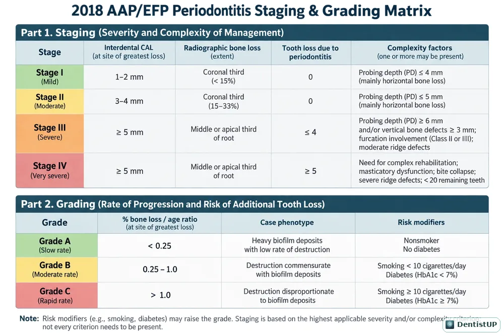
Stage is based on the site of greatest loss. Work through three measures, then check complexity:
- Stage I: interdental CAL 1–2 mm; radiographic bone loss in the coronal third (under 15%); no tooth loss due to periodontitis; maximum probing depth 4 mm or less, mostly horizontal bone loss.
- Stage II: CAL 3–4 mm; bone loss in the coronal third (15–33%); no tooth loss due to periodontitis; maximum probing depth 5 mm or less, mostly horizontal bone loss.
- Stage III: CAL 5 mm or more; bone loss extending to the middle or apical third of the root; tooth loss due to periodontitis of 4 teeth or fewer. Complexity may include probing depths of 6 mm or more, vertical bone loss of 3 mm or more, Class II or III furcation involvement and a moderate ridge defect.
- Stage IV: as Stage III for CAL and bone loss, but tooth loss due to periodontitis of 5 teeth or more, and a need for complex rehabilitation because of masticatory dysfunction, secondary occlusal trauma (tooth mobility degree 2 or more), a severe ridge defect, bite collapse, drifting or flaring, or fewer than 20 remaining teeth (10 opposing pairs).
Two rules settle most exam questions. First, if CAL is unavailable, use radiographic bone loss. Second, a single complexity factor can shift the stage upwards: a patient with Stage II attachment loss but a Class II furcation is Stage III.
Step 3: Extent
Describe how widespread the disease is: localised when fewer than 30% of teeth are involved, generalised when 30% or more are involved, or a molar/incisor pattern, which replaces much of what used to be called localised aggressive periodontitis.
Step 4: Grade (rate of progression)
Start at Grade B, then look for evidence to move up or down.
- Direct evidence: radiographic bone loss or CAL change over five years. No loss suggests Grade A, less than 2 mm suggests Grade B, and 2 mm or more suggests Grade C.
- Indirect evidence: percentage of bone loss at the worst tooth divided by the patient's age. Below 0.25 suggests Grade A, 0.25 to 1.0 suggests Grade B, and above 1.0 suggests Grade C. The case phenotype also matters: heavy biofilm with little destruction points to Grade A; destruction out of proportion to biofilm points to Grade C.
- Grade modifiers: smoking and diabetes. Fewer than 10 cigarettes a day or HbA1c below 7% in a patient with diabetes supports Grade B; 10 or more cigarettes a day or HbA1c of 7% or higher supports Grade C.
Worked examples
Example 1. A 35-year-old non-smoker has interdental CAL of 5 mm on the mesial of 36 and 46, bone loss to the middle third of the roots, no missing teeth and no furcation involvement. Six of 28 teeth are affected. Bone loss at the worst site is about 40%, so the ratio is 40 ÷ 35 ≈ 1.1. Diagnosis: periodontitis, Stage III, localised, Grade C. The trap is to call it Stage II because nothing looks complex; the 5 mm CAL decides the stage.
Example 2. A 60-year-old smoking 5 cigarettes a day has generalised CAL of 3–4 mm with horizontal bone loss of about 20% and no teeth lost to periodontitis. The ratio is 20 ÷ 60 ≈ 0.33. Diagnosis: periodontitis, Stage II, generalised, Grade B. Light smoking supports Grade B; it would take 10 or more a day to reach Grade C.
Example 3. A patient with Stage III findings has lost six teeth to periodontitis, has mobility of degree 2 on several teeth and has 18 remaining teeth. Diagnosis: Stage IV, because both tooth loss and the need for complex rehabilitation exceed Stage III.
Common traps
- Staging on the average rather than the worst site.
- Counting all missing teeth instead of only those lost to periodontitis.
- Forgetting that one complexity factor, such as a Class II furcation, can raise the stage.
- Lowering the stage after successful treatment. Stage does not go down.
- Calling smoking "a risk factor" without checking the number of cigarettes a day, which changes the grade.
Beyond periodontitis
The same classification defines periodontal health, gingivitis and peri-implant conditions. On an intact periodontium, gingivitis is generally defined by bleeding on probing at 10% or more of sites with probing depths of 3 mm or less. Peri-implant mucositis shows inflammation without progressive bone loss, while peri-implantitis includes progressive bone loss; a history of periodontitis and poor plaque control raise the risk.
Question 2 · Periodontics · Peri-implant Disease
Which history increases a patient's risk of peri-implantitis?
- AAdequate cleansability around a well-designed restoration
- BA stable baseline radiograph after physiologic remodeling
- CPrevious periodontitis combined with poor plaque control
- DConsistently healthy tissues and reliable supportive maintenance
Show answer and explanation
Answer: C. Previous periodontitis combined with poor plaque control
A history of periodontitis and poor plaque control are important peri-implantitis risk indicators; smoking, uncontrolled diabetes, and lack of maintenance can add risk. Stable health, accessible prosthetic design, and supportive care are favorable. Risk history prompts prevention and monitoring rather than proving current disease.
Concept to remember: A history of periodontitis and poor plaque control are key risk indicators for peri-implantitis.
Practise with the free periodontics questions, and see the Visual Atlas for the biologic width and peri-implant disease illustrations.
Frequently asked questions
What is the difference between staging and grading?+
Staging describes how severe and complex the disease is now, based mainly on attachment loss, bone loss, tooth loss and complexity factors. Grading describes the rate of progression and risk of future progression, using direct or indirect evidence of progression and risk factors such as smoking and diabetes.
Which grade should I assume if there is no evidence of progression rate?+
Grade B, moderate rate of progression, is the default. You then move to Grade A or C only when there is evidence to support it.
How do smoking and diabetes change the grade?+
Under the 2017 framework, smoking fewer than 10 cigarettes a day or diabetes with HbA1c below 7% supports Grade B; smoking 10 or more cigarettes a day or HbA1c of 7% or higher supports Grade C.
When is periodontitis localised or generalised?+
Localised when fewer than 30% of teeth are involved and generalised when 30% or more are involved. A molar/incisor pattern is described separately.
Can the stage go down after treatment?+
No. Stage is determined by the worst attachment and bone loss the patient has experienced and does not decrease after successful treatment. Grade can be reassessed.
Free practice
DentistUP is an independent educational platform. Socket Science is not affiliated with, endorsed by, sponsored by, or approved by NDEB, INBDE, JCNDE, ADA, GDC, ORE, ADC, or any other examination authority. DentistUP does not contain official or recalled examination questions and does not guarantee examination outcomes.