SocketScience

New in DentistUP 2.1

The Visual Atlas: 260+ dental illustrations

Anatomy diagrams, clinical illustrations and comparison charts, built into the topic lessons and collected in one place. Search the atlas, filter by branch, save favourites, and open the lesson a visual belongs to.

260+

illustrations

263

illustrated lessons

29

branches

Where the visuals appear in the app

  • Inside every lesson

    Diagrams and charts sit next to the text they explain.

  • Related visuals

    Lessons suggest visuals from other topics. The impacted third molar lesson links to mandible, trigeminal nerve and maxillary sinus diagrams. Open the source topic in a quick-view sheet and come back to the same spot.

  • Under question explanations

    Each explanation shows the illustrations for that topic.

  • In the Visual Atlas

    Every illustration in one searchable place, filterable by branch, with favourites.

A look inside: 24 examples by branch

A small sample. The app holds 260+ illustrations across all 29 branches.

Periodontics
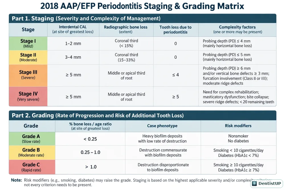
Chart of the 2018 AAP/EFP periodontitis staging (Stage I to IV) and grading (Grade A to C) criteria
Periodontitis staging and grading matrix
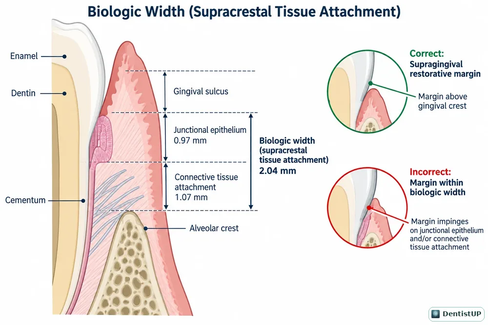
Diagram of biologic width: sulcus depth, junctional epithelium and connective tissue attachment
Supracrestal tissue attachment

Tooth Anatomy & Morphology

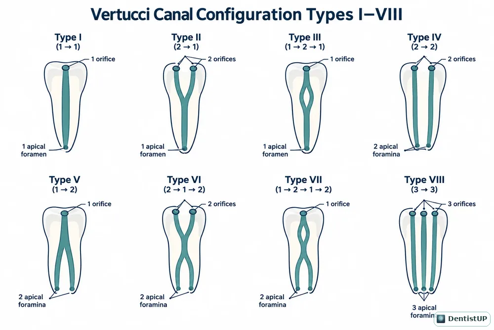
Diagram of Vertucci root canal configuration types I to VIII
Vertucci canal configurations I–VIII
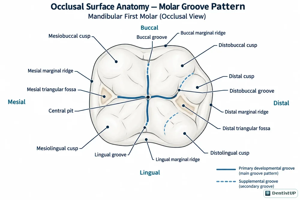
Occlusal view of a mandibular first molar with cusps, ridges, fossae and grooves labelled
Mandibular first molar occlusal anatomy

Head & Neck Anatomy

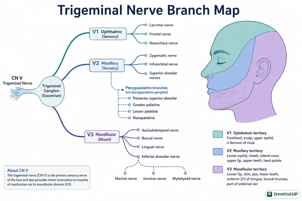
Branch map of the trigeminal nerve showing V1, V2 and V3 divisions and their territories
Trigeminal nerve branch map
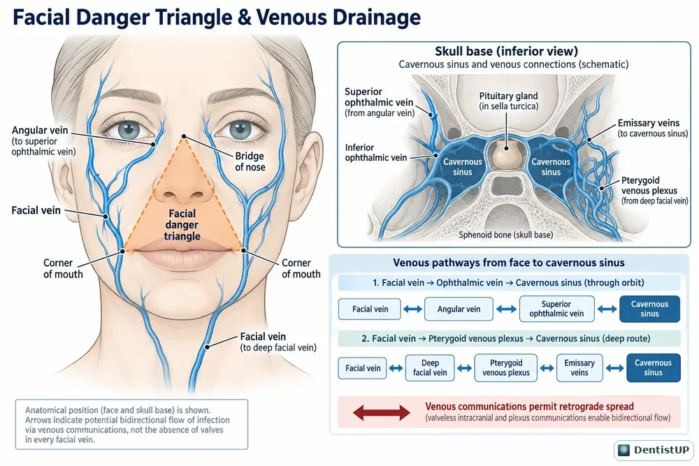
Facial danger triangle with venous pathways to the cavernous sinus
Facial danger triangle and venous drainage
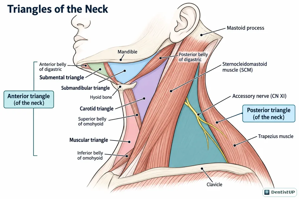
Anterior and posterior triangles of the neck with their subdivisions
Triangles of the neck
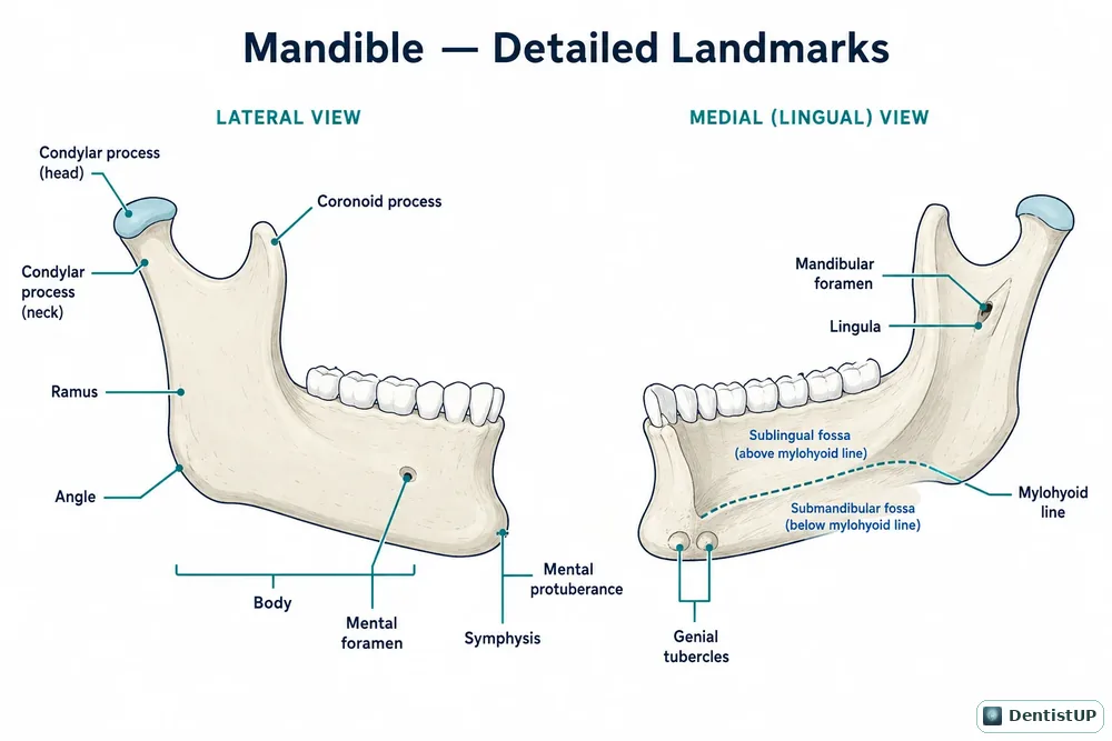
Lateral and medial views of the mandible with landmarks labelled
Mandible: detailed landmarks

Endodontics

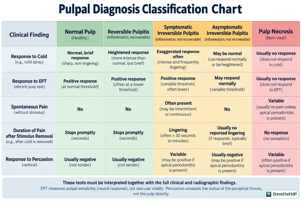
Chart comparing normal pulp, reversible pulpitis, irreversible pulpitis and pulp necrosis by clinical test findings
Pulpal diagnosis classification
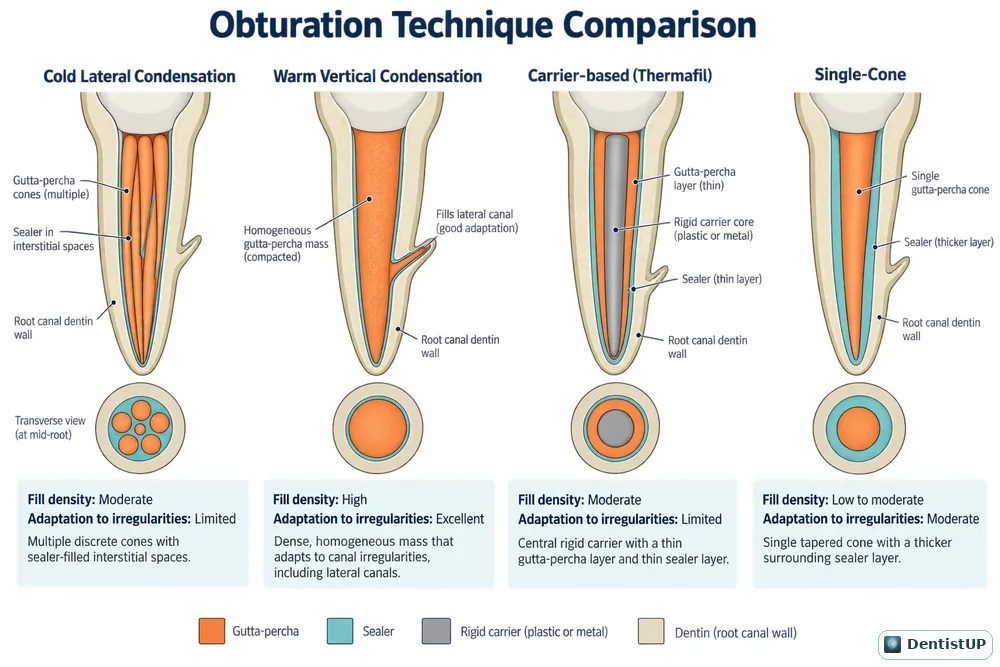
Comparison of root canal obturation techniques and their fill density
Obturation techniques compared

Oral & Maxillofacial Surgery

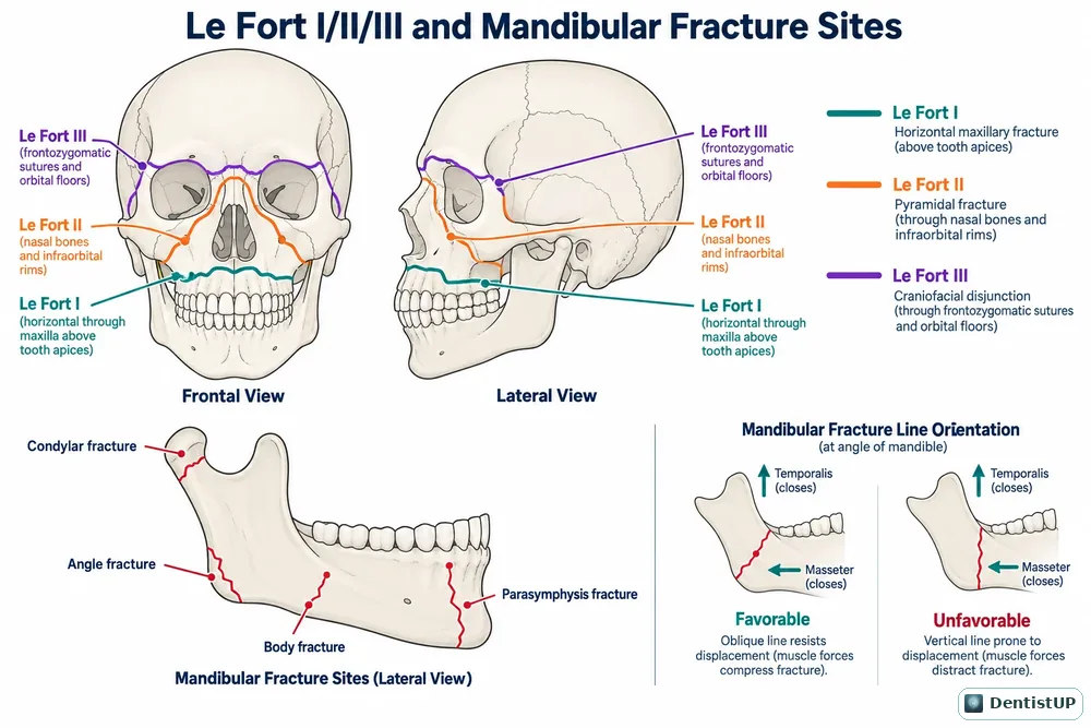
Frontal and lateral skull diagrams showing Le Fort I, II and III fracture lines and mandibular fracture sites
Le Fort I/II/III and mandibular fracture sites
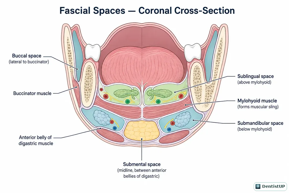
Coronal cross-section showing buccal, sublingual, submandibular and submental fascial spaces
Fascial spaces in coronal section
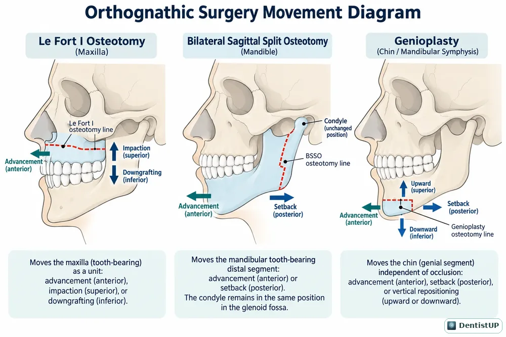
Le Fort I osteotomy, bilateral sagittal split osteotomy and genioplasty movement diagrams
Orthognathic surgery movements

Oral Pathology

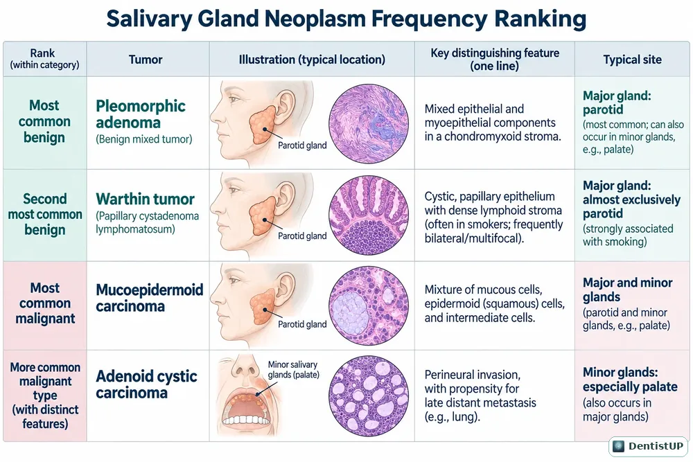
Table ranking pleomorphic adenoma, Warthin tumour, mucoepidermoid carcinoma and adenoid cystic carcinoma by frequency
Salivary gland neoplasm frequency ranking
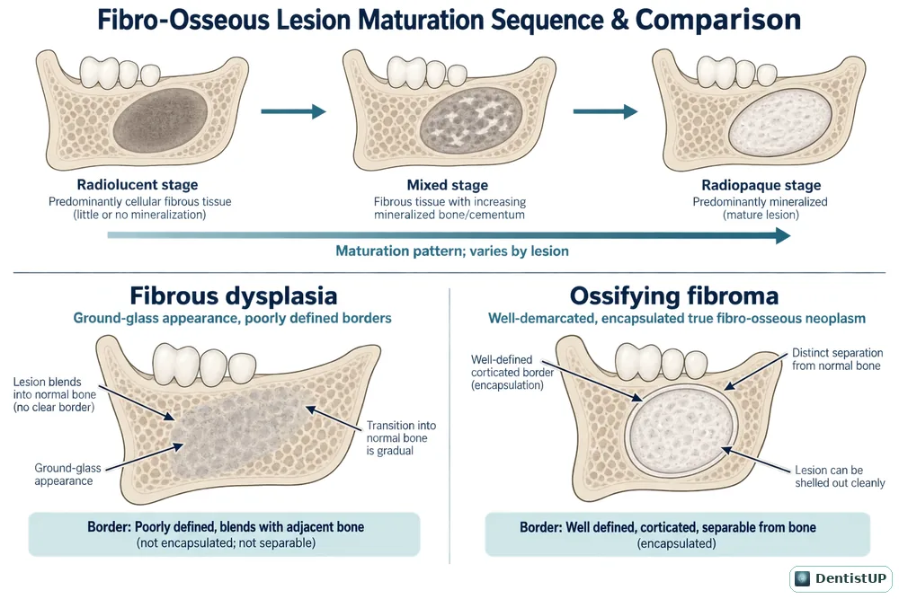
Comparison of fibrous dysplasia and ossifying fibroma borders and radiographic maturation stages
Fibro-osseous lesions compared
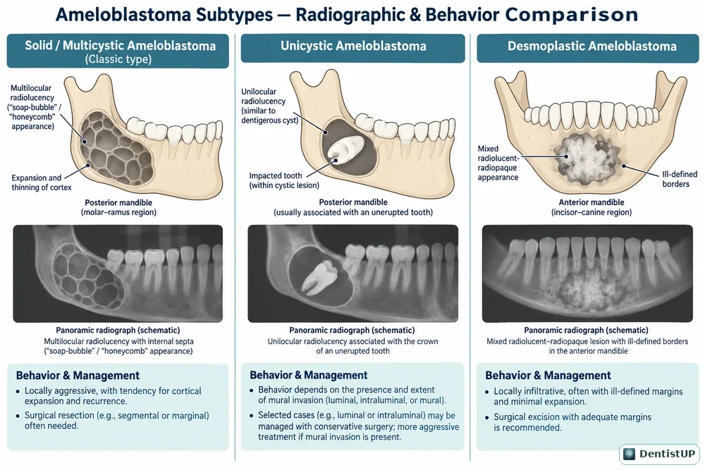
Solid, unicystic and desmoplastic ameloblastoma radiographic and behavioural comparison
Ameloblastoma subtypes on radiographs

Anesthesia & Pain Control

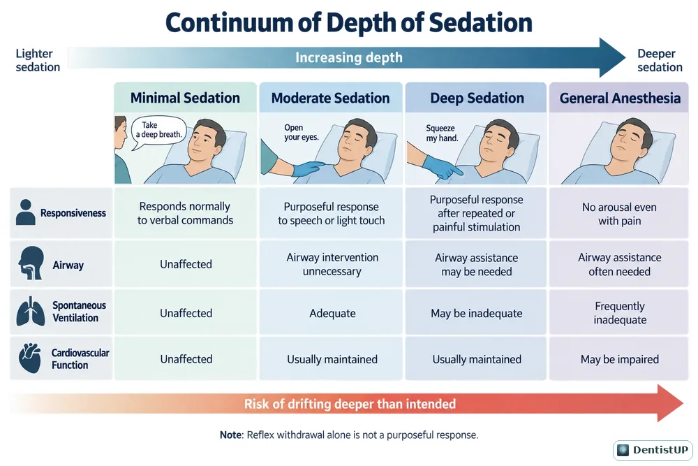
Chart comparing minimal, moderate and deep sedation and general anaesthesia
Continuum of depth of sedation

Oral Medicine

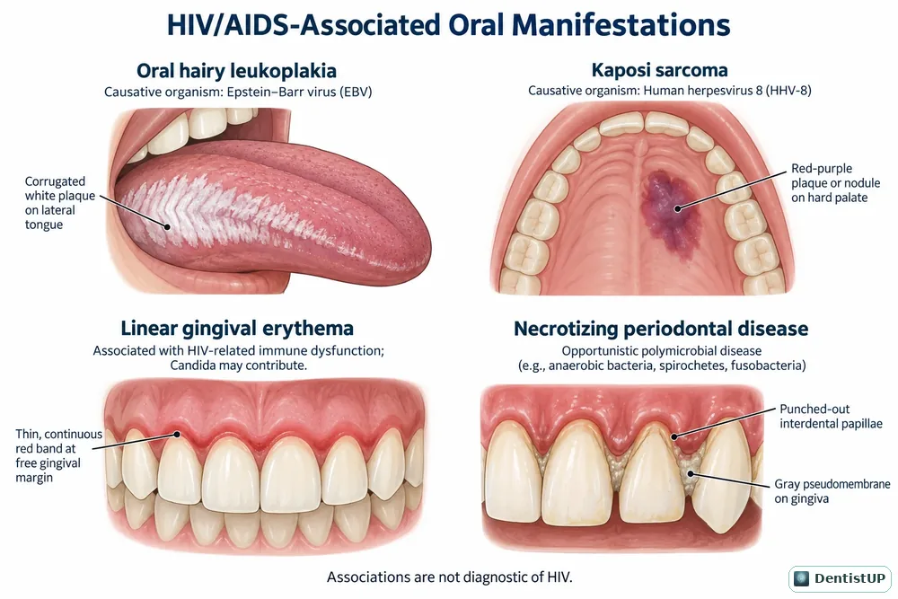
Illustrations of oral hairy leukoplakia, Kaposi sarcoma, linear gingival erythema and necrotising periodontal disease
HIV-associated oral manifestations

Histology & Embryology

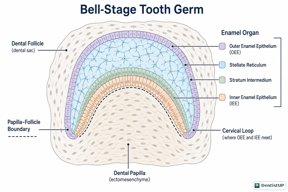
Histological illustration of a tooth germ at the bell stage of odontogenesis
Tooth germ at the bell stage
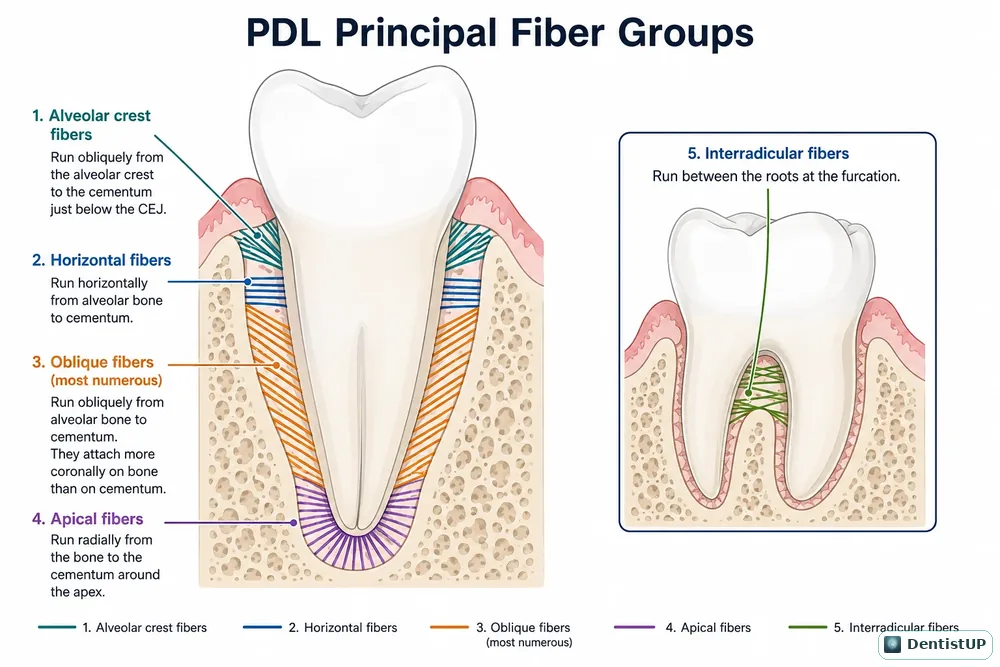
Diagram of the principal periodontal ligament fibre groups
Periodontal ligament fibre groups

Pediatric Dentistry

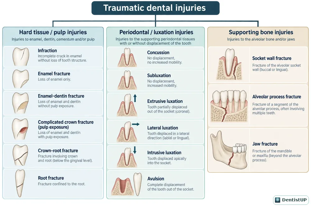
Chart classifying dental traumatic injuries in children
Dental trauma classification

Biochemistry

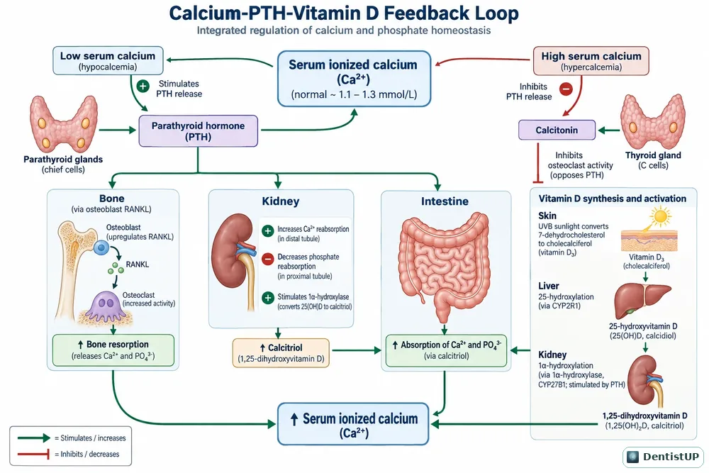
Feedback loop between serum calcium, parathyroid hormone and vitamin D across bone, kidney and intestine
Calcium, PTH and vitamin D feedback loop

Evidence-Based Dentistry & Biostatistics
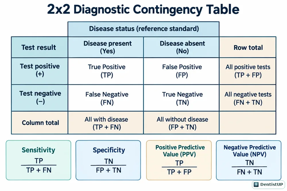
2x2 table with sensitivity, specificity, positive and negative predictive value formulas
2×2 diagnostic contingency table

Infection Control

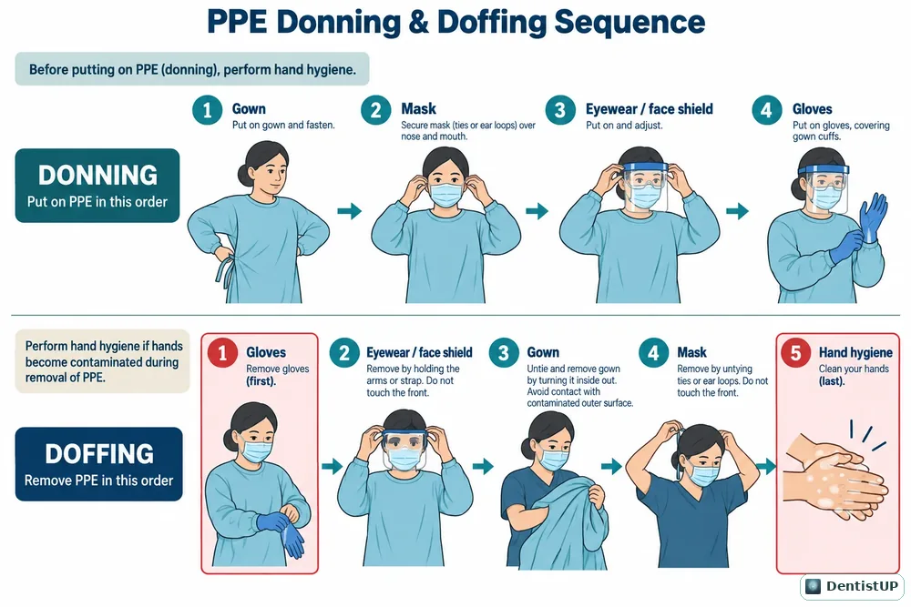
Step-by-step sequence for putting on and removing personal protective equipment
PPE donning and doffing sequence

Illustrations © Socket Science. For personal study; please do not republish. Educational material, not patient-specific clinical guidance.