
Impacted Third Molars for Dental Boards: Anatomy, Winter's Classification and Nerve Risk
Impacted third molars bring together anatomy, radiology, surgery and consent, which is why they appear on every dental board exam. This guide covers the classifications you need, the radiographic signs of nerve risk, and the anatomy that explains them.
By the Socket Science editorial team · Reviewed by a qualified dentist
Independent resource. DentistUP is not affiliated with or endorsed by any examination authority. Always check the official exam body for current format, dates and eligibility. Educational content, not patient-specific clinical advice.
Why third molars get impacted
A tooth is impacted when it fails to erupt into its normal functional position within the expected time, usually because it is blocked by another tooth, bone or soft tissue. Mandibular third molars are the most frequently impacted teeth. Exam questions on them fall into four groups: classification, assessment of nerve risk, indications for removal, and management of complications. Each depends on the anatomy of the posterior mandible, so start there.
The anatomy that matters
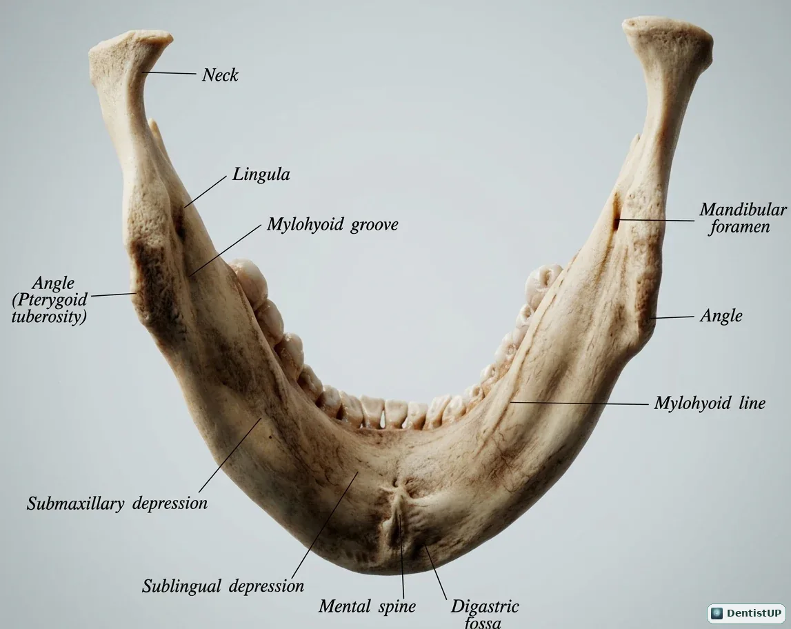
The inferior alveolar nerve enters the mandibular foramen on the medial surface of the ramus, runs forward in the mandibular canal below the molar roots, and gives off the mental nerve at the mental foramen. Its relationship to the third molar roots is the single most important factor in surgical risk.
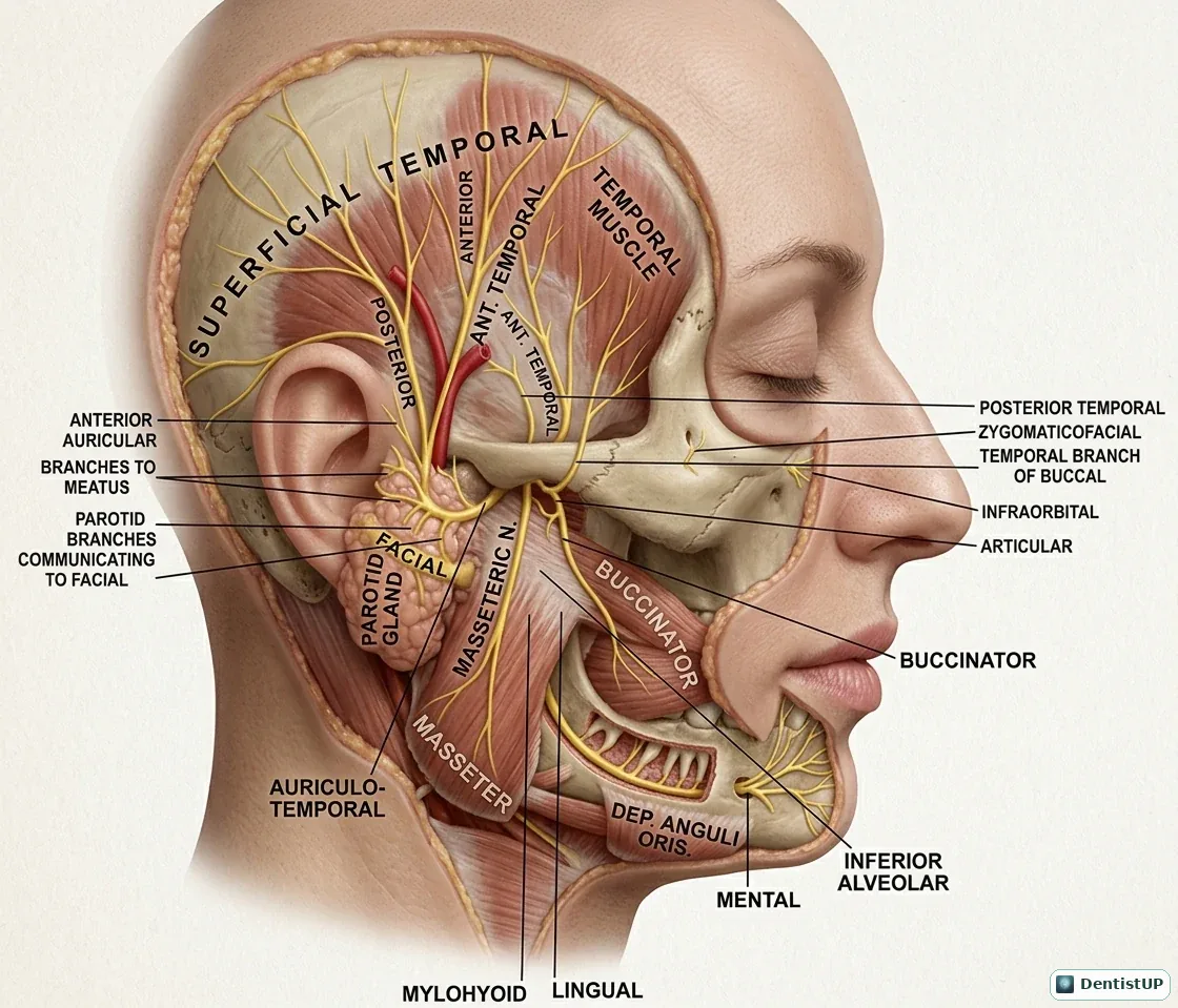
The lingual nerve, a branch of the mandibular division of the trigeminal nerve, runs forward close to the lingual cortical plate in the third molar region. In some patients it lies at or even above the level of the alveolar crest. Lingual flap retraction and distolingual bone removal put it at risk, and injury causes altered sensation of the anterior two-thirds of the tongue, often with altered taste because the chorda tympani fibres travel with it.
The mylohyoid line on the medial surface also matters. Third molar apices usually lie below it, so infection from these teeth tends to spread into the submandibular space rather than the sublingual space.
Winter's classification: angulation
Winter's classification describes the angle of the third molar's long axis relative to that of the second molar:
- Mesioangular: the crown tilts forward towards the second molar. Generally reported as the most common impaction.
- Vertical: the long axis is roughly parallel to the second molar.
- Horizontal: the long axis is roughly perpendicular to the second molar.
- Distoangular: the crown tilts back towards the ramus. In the mandible this is usually considered the most difficult to remove, because the path of removal runs into the ramus.
Transverse (buccoangular or linguoangular) and inverted positions also occur but are less commonly tested. Note that difficulty rankings differ between the mandible and maxilla: in the maxilla, the distoangular position is usually the easier one.
Pell and Gregory: space and depth
Pell and Gregory classify two separate things.
Relationship to the ramus (space):
- Class I: enough space between the anterior border of the ramus and the distal surface of the second molar for the whole mesiodistal width of the third molar crown.
- Class II: less space than the width of the crown; part of the crown lies within the ramus.
- Class III: all or most of the crown lies within the ramus.
Depth relative to the second molar:
- Position A: the highest point of the third molar is at or above the occlusal plane of the second molar.
- Position B: the highest point is below the occlusal plane but above the cervical line of the second molar.
- Position C: the highest point is below the cervical line of the second molar.
Difficulty rises from Class I to Class III and from Position A to Position C. Exam questions often give a description and ask for the classification, or give two classifications and ask which extraction is likely to be more difficult.
Radiographic signs of nerve proximity
On a panoramic radiograph, several classic signs suggest that the roots and the inferior alveolar canal are closely related. They are often attributed to Rood and Shehab:
- Darkening of the root where the canal crosses it, the sign most often associated with nerve injury risk.
- Interruption of the white line (the radiopaque borders) of the canal.
- Diversion of the canal, with the canal bending around the roots.
- Narrowing of the canal as it crosses the roots.
- Deflection or narrowing of the roots, and a dark, bifid apex.
A panoramic image is two-dimensional, so these signs indicate risk rather than prove contact. When they are present, three-dimensional imaging such as CBCT is often considered to show whether the canal lies buccal, lingual or between the roots, which informs both consent and technique.
Indications for removal
Board exams generally reward removal for a clear reason rather than routine prophylactic removal of asymptomatic, disease-free teeth. Commonly cited indications include recurrent or severe pericoronitis, unrestorable caries in the third molar, caries or resorption of the adjacent second molar attributable to the third molar, periodontal disease affecting the distal of the second molar, cysts or tumours associated with the tooth, and surgical need, for example before orthognathic surgery or in a fracture line. Local guidelines differ by country, so check the guidance relevant to your exam.
Reducing and managing nerve risk
- Assess and consent. Document radiographic signs, discuss the risk of temporary and permanent altered sensation, and discuss alternatives including monitoring.
- Protect the lingual side. Careful flap design and retraction, and cautious bone removal distolingually.
- Section the tooth to reduce the force and bone removal needed.
- Consider coronectomy when roots are intimately related to the canal: the crown is removed and the roots are deliberately left in place.
- Refer when the case exceeds your competence, a principle that also appears in ethics questions.
Other complications worth knowing
Beyond nerve injury, expect questions on alveolar osteitis (dry socket), usually presenting a few days after extraction with increasing pain and an empty socket; trismus and swelling; bleeding, especially in patients on anticoagulants or antiplatelets; infection, including spread to the submandibular and pterygomandibular spaces; and, for maxillary third molars, displacement into the maxillary sinus or infratemporal fossa and oroantral communication.
Keep going
Test yourself with the free oral and maxillofacial surgery questions, including a Pell and Gregory question, and the head and neck anatomy questions. For the anatomy, the submandibular and sublingual spaces diagram explains where infection spreads, and our guide to local anaesthesia for dental boards covers the inferior alveolar nerve block.
Frequently asked questions
What is the most common angulation of an impacted mandibular third molar?+
Mesioangular impaction is generally reported as the most common. Distoangular impactions of the mandibular third molar are usually considered the most difficult to remove.
What does Pell and Gregory Class I mean?+
There is enough space between the anterior border of the ramus and the distal surface of the second molar to accommodate the mesiodistal width of the third molar crown. Class II has partial space, and in Class III the crown lies largely within the ramus.
Which radiographic signs suggest the inferior alveolar canal is close to the roots?+
Classic panoramic signs include darkening of the root where the canal crosses it, interruption of the canal's white line, diversion of the canal, and narrowing of the canal or the root. When present, three-dimensional imaging such as CBCT is often considered.
Which nerve is at risk on the lingual side?+
The lingual nerve. It can lie close to the lingual cortical plate in the third molar region, and occasionally at or above the crest, so lingual retraction and distolingual bone removal carry risk of injury.
What is coronectomy?+
Removal of the crown of a third molar while deliberately leaving the roots in place, used in selected cases where the roots are intimately related to the inferior alveolar canal, to reduce the risk of nerve injury.
Free practice
DentistUP is an independent educational platform. Socket Science is not affiliated with, endorsed by, sponsored by, or approved by NDEB, INBDE, JCNDE, ADA, GDC, ORE, ADC, or any other examination authority. DentistUP does not contain official or recalled examination questions and does not guarantee examination outcomes.