
Head & Neck Anatomy Practice Questions
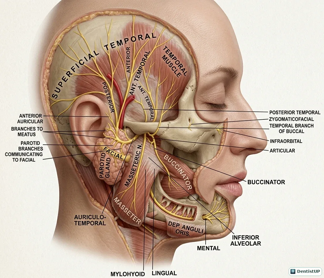
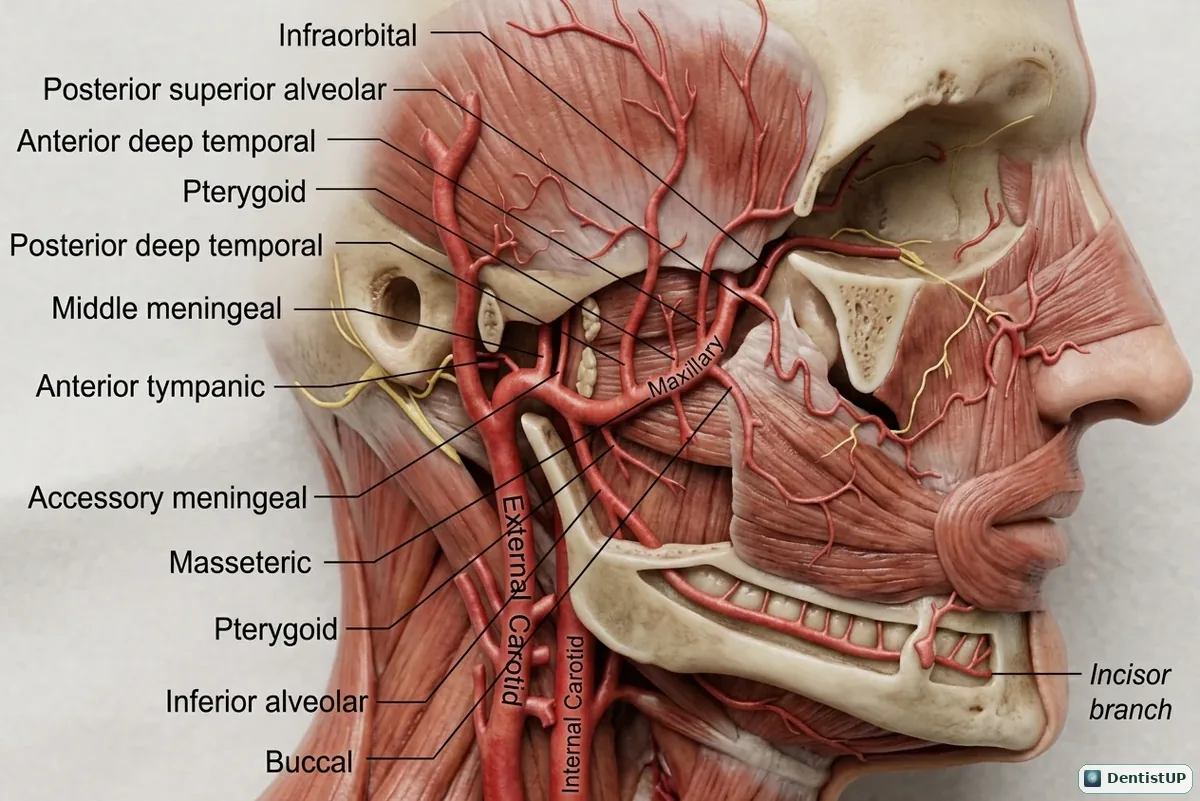
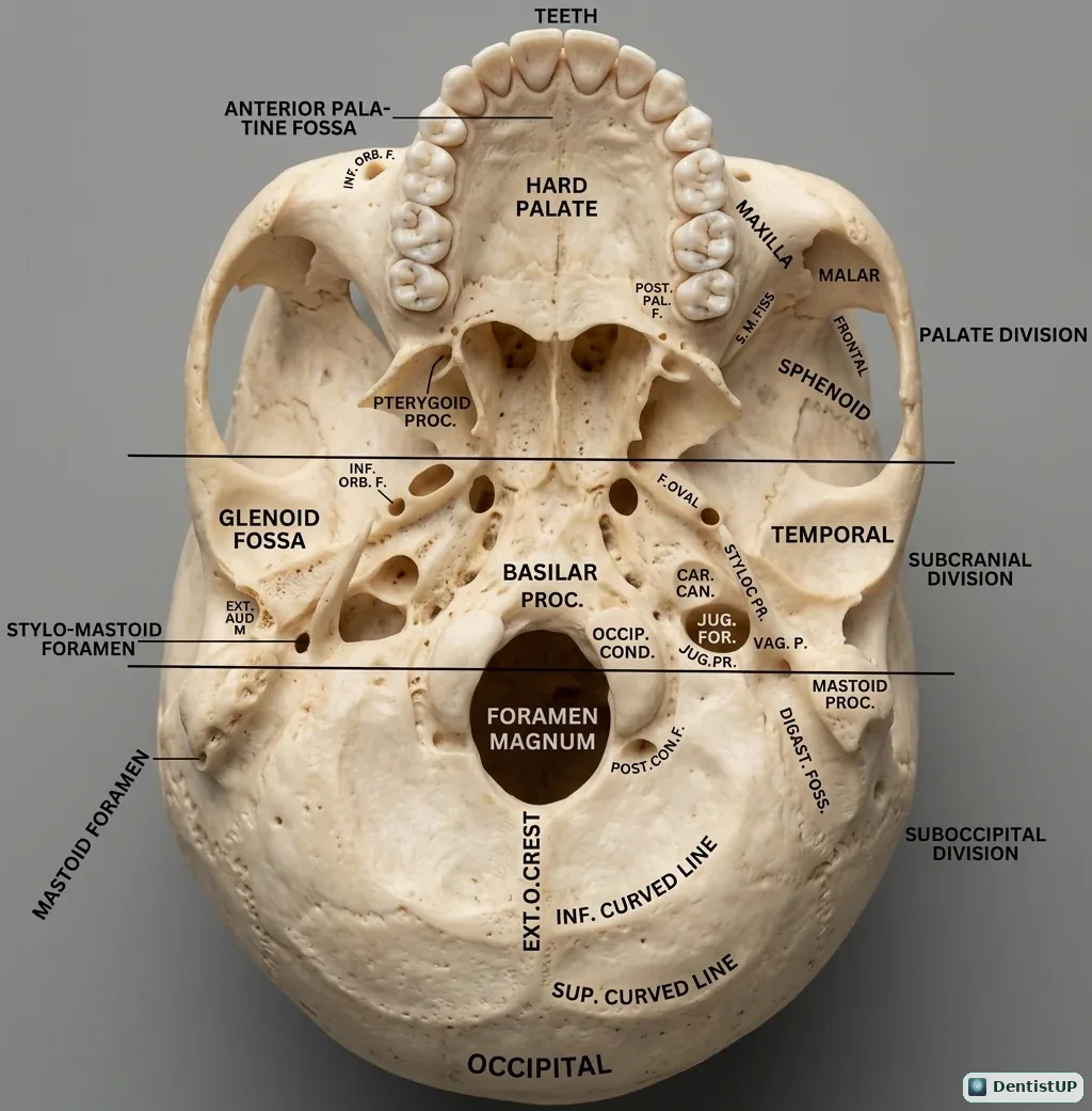
Head and neck anatomy underpins local anaesthesia, oral surgery, radiology and the spread of infection, which is why it runs through every dental board exam and through the first years of dental school. These questions cover skeletal anatomy, cranial foramina and their contents, muscles, innervation, vasculature, fascial spaces, paranasal sinuses, the TMJ, the pharynx, larynx and airway, and cephalometric and radiographic landmarks. They pair well with our free head and neck anatomy diagrams, because most anatomy questions are easier once you can picture the structure and its relations.
10 questions · answers and explanations revealed as you go · no sign-up
Questions
Question 1 of 10
Paranasal Sinuses
The sphenoid sinus is located within which bone?
The answer is D.
The sphenoid sinus is located within the body of the sphenoid bone, positioned deep within the skull near midline structures such as the pituitary gland, internal carotid artery, and optic nerve, making its disease potentially serious.
Question 2 of 10
Muscles
The orbicularis oris muscle is primarily responsible for which action?
The answer is C.
The orbicularis oris is a circular sphincter muscle surrounding the mouth that closes and purses the lips, playing an important role in speech, feeding, and maintaining oral competence, and it receives its motor supply from the buccal and marginal mandibular branches of the facial nerve.
Question 3 of 10
Vasculature
The occipital artery, a branch of the external carotid artery, primarily supplies which region?
The answer is A.
The occipital artery branches from the external carotid artery and travels posteriorly to supply the scalp over the back of the skull, running a course that is relevant to surgical approaches involving the posterior neck and occipital region.
Question 4 of 10
TMJ
The stylomandibular ligament connects the styloid process to which part of the mandible?
The answer is D.
The stylomandibular ligament is a band of deep cervical fascia that extends from the styloid process of the temporal bone to the angle and posterior border of the mandibular ramus, helping to limit excessive protrusion of the mandible.
Question 5 of 10
Fascial Spaces
The submental space is located in which anatomic position?
The answer is A.
The submental space lies below the chin in the midline, bounded by the mylohyoid muscle above, the anterior bellies of the digastric muscles laterally, and the overlying skin and superficial fascia, and it is a common site of swelling from anterior mandibular incisor infections.
Question 6 of 10
Cranial Foramina & Contents
The foramen rotundum primarily transmits which structure?
The answer is B.
The foramen rotundum is a round opening in the greater wing of the sphenoid bone that transmits the maxillary division of the trigeminal nerve (V2) from the middle cranial fossa into the pterygopalatine fossa, on its way to supplying the midface and upper teeth.
Question 7 of 10
Innervation
The accessory nerve (cranial nerve XI) is clinically significant during neck surgery mainly because it supplies which muscles?
The answer is D.
The accessory nerve supplies the sternocleidomastoid and trapezius muscles, and because it travels a relatively superficial course through the posterior triangle of the neck, it is at recognized risk of injury during lymph node dissection or biopsy in that region.
Question 8 of 10
Cephalometric/Radiographic Landmarks
The cephalometric point Gonion represents which anatomic structure?
The answer is D.
Gonion is defined as the point at the angle of the mandible where the ramus and body meet, and its position helps define the gonial angle, a measurement used to assess mandibular growth pattern and facial type.
Question 9 of 10
Skeletal Anatomy
The genial tubercles on the internal surface of the mandibular symphysis primarily serve which function?
The answer is D.
The genial tubercles are small bony projections on the internal surface of the mandibular symphysis that serve as the attachment sites for the genioglossus muscle superiorly and the geniohyoid muscle inferiorly, both important for tongue position and airway support.
Question 10 of 10
Pharynx, Larynx & Airway
The laryngopharynx extends from the epiglottis down to which landmark?
The answer is C.
The laryngopharynx, also called the hypopharynx, extends from the upper border of the epiglottis down to the lower border of the cricoid cartilage, where it becomes continuous with the esophagus, and it surrounds the entrance to the larynx.
Choose an answer to reveal the explanation.
Related anatomy diagrams
More branches
Full mock exams
DentistUP is an independent educational platform. Socket Science is not affiliated with, endorsed by, sponsored by, or approved by NDEB, INBDE, JCNDE, ADA, GDC, ORE, ADC, or any other examination authority. DentistUP does not contain official or recalled examination questions and does not guarantee examination outcomes. Educational content, not patient-specific clinical advice.