
Skeletal Anatomy
Medial Surface of the Mandible
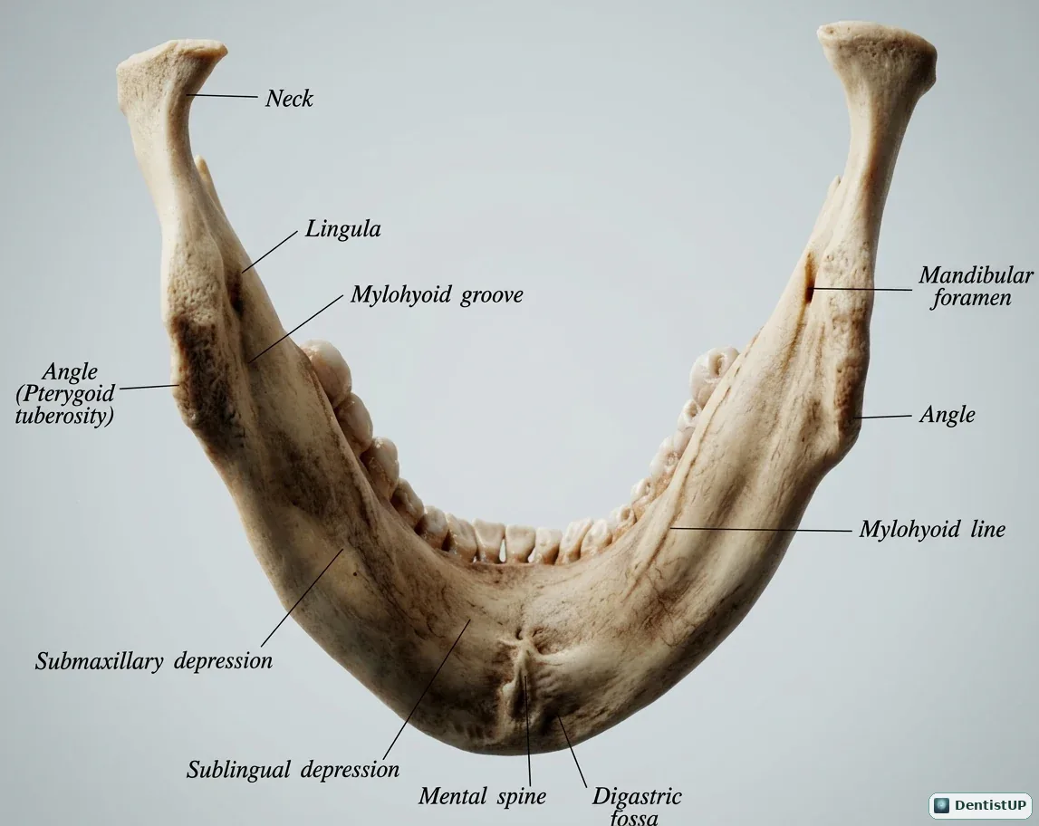
The medial surface of the mandible carries the landmarks that matter most for local anaesthesia and oral surgery. This illustration shows the mandibular foramen and the lingula that overlaps it, the mylohyoid groove and mylohyoid line, the sublingual and submandibular fossae, the genial tubercles (mental spines) and the digastric fossa.
Exam questions use these landmarks in three ways. The mandibular foramen and lingula explain where the inferior alveolar nerve block deposits solution and why it can fail. The mylohyoid line divides the sublingual from the submandibular space, which determines where an infection from a lower molar spreads. The genial tubercles give attachment to genioglossus and geniohyoid, a common anatomy recall item.
Related practice questions
Educational illustration for exam preparation, not patient-specific clinical guidance.