
Fascial Spaces
Submandibular and Sublingual Spaces
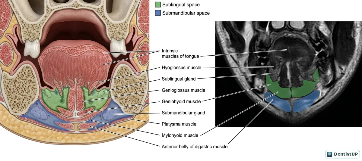
A coronal section through the floor of the mouth, paired with a matching cross-sectional image. It shows the sublingual space above mylohyoid, containing the sublingual gland, and the submandibular space below it, along with the intrinsic tongue muscles, hyoglossus, genioglossus, geniohyoid, platysma and the anterior belly of digastric.
Fascial spaces are a high-yield oral surgery topic. The rule to remember is that mylohyoid decides the route: infection from teeth whose apices lie above the mylohyoid line (premolars and first molar) enters the sublingual space, while infection from the second and third molars, whose apices lie below it, enters the submandibular space. Bilateral involvement of the submandibular, sublingual and submental spaces is Ludwig's angina, an airway emergency.
Related practice questions
Educational illustration for exam preparation, not patient-specific clinical guidance.