
Neck Anatomy
Deep Cervical Fascia Layers
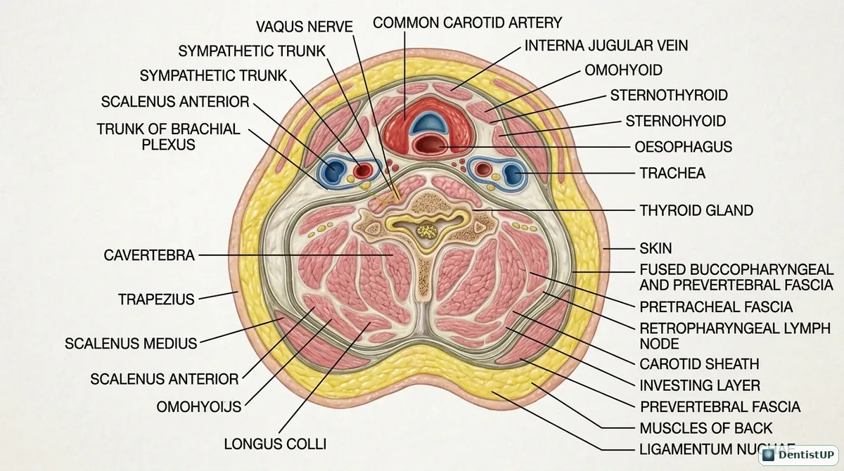
An axial section of the neck showing the layers of deep cervical fascia: the investing layer surrounding sternocleidomastoid and trapezius, the pretracheal (visceral) layer around the thyroid, trachea and oesophagus, the prevertebral layer around the vertebral muscles, and the carotid sheath containing the carotid artery, internal jugular vein and vagus nerve.
Fascial layers explain how odontogenic infections become life-threatening. Infection spreading from the parapharyngeal space can enter the retropharyngeal space, and from there the danger space between the alar and prevertebral fascia, which runs down into the mediastinum. Exam questions ask which space lies in this pathway and why a patient with a lower molar infection and difficulty swallowing needs urgent assessment.
Related practice questions
Educational illustration for exam preparation, not patient-specific clinical guidance.