
Neck Anatomy
Anterior Neck Triangles
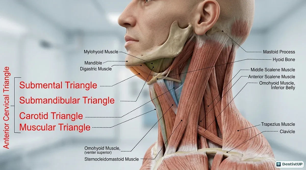
A lateral view of the neck dividing the anterior cervical triangle into the submental, submandibular, carotid and muscular triangles, with mylohyoid, digastric, sternocleidomastoid, omohyoid and trapezius as boundaries.
The neck triangles organise lymph node examination and the spread of infection. Submental nodes drain the lower incisors, lower lip and tip of the tongue; submandibular nodes drain most other teeth and much of the tongue, so an enlarged node here often points to a dental cause. The carotid triangle contains the carotid bifurcation, internal jugular vein and vagus nerve, which is where the carotid pulse is felt during an emergency. The muscular triangle holds the infrahyoid muscles over the thyroid gland and larynx.
Related practice questions
Educational illustration for exam preparation, not patient-specific clinical guidance.