
Skeletal Anatomy
Lateral Skull Landmarks
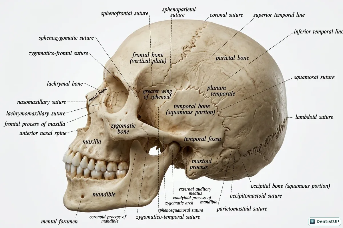
This lateral view of the skull labels the major sutures (coronal, squamous, lambdoid, sphenoparietal and sphenosquamous), the temporal lines and temporal fossa, the zygomatic arch, the external acoustic meatus, the mastoid and styloid processes, and the mandible with its coronoid and condylar processes.
For dental board exams the lateral skull is the map behind several applied topics: the temporal fossa and zygomatic arch frame the path of temporalis, the articular eminence and glenoid fossa define TMJ movement, and the pterion marks the thin area overlying the middle meningeal artery. The same landmarks reappear on lateral cephalograms and panoramic radiographs, so recognising them here makes radiology questions easier.
Related practice questions
Educational illustration for exam preparation, not patient-specific clinical guidance.