
Vasculature
Head and Neck Venous Drainage
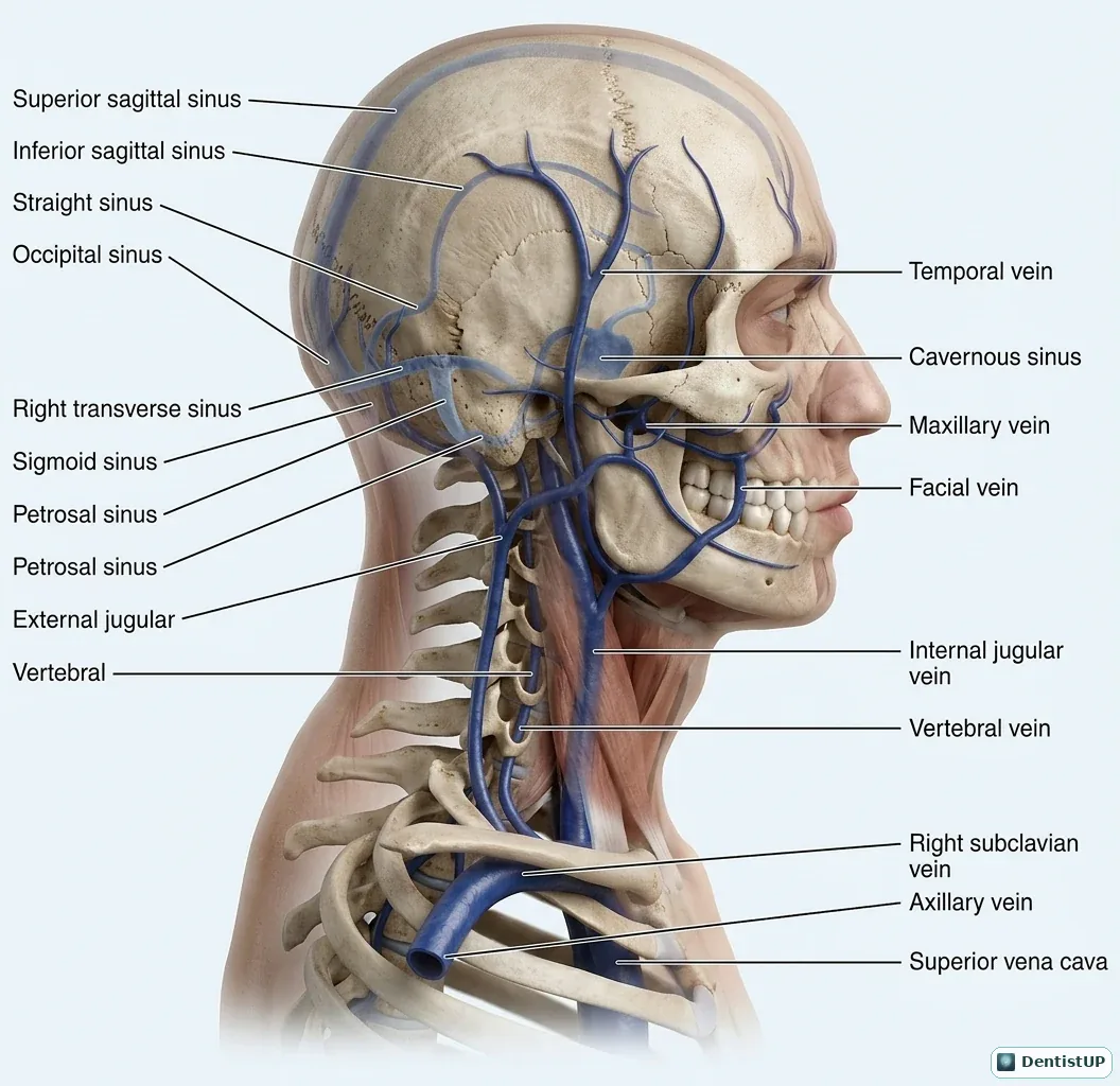
This illustration traces venous drainage from the dural venous sinuses (superior and inferior sagittal, straight, transverse, sigmoid and petrosal) and from the face (facial, maxillary and temporal veins) into the internal and external jugular veins, the subclavian vein and the superior vena cava.
The link most often tested is between the face and the cavernous sinus. The facial vein connects with the ophthalmic veins and, through the pterygoid plexus, with the cavernous sinus; these veins have few effective valves. That is why an infection in the danger triangle of the face or from maxillary teeth can, rarely, lead to cavernous sinus thrombosis. The pterygoid plexus also explains haematoma after a posterior superior alveolar block.
Related practice questions
Educational illustration for exam preparation, not patient-specific clinical guidance.