
Vasculature
Maxillary Artery Branches
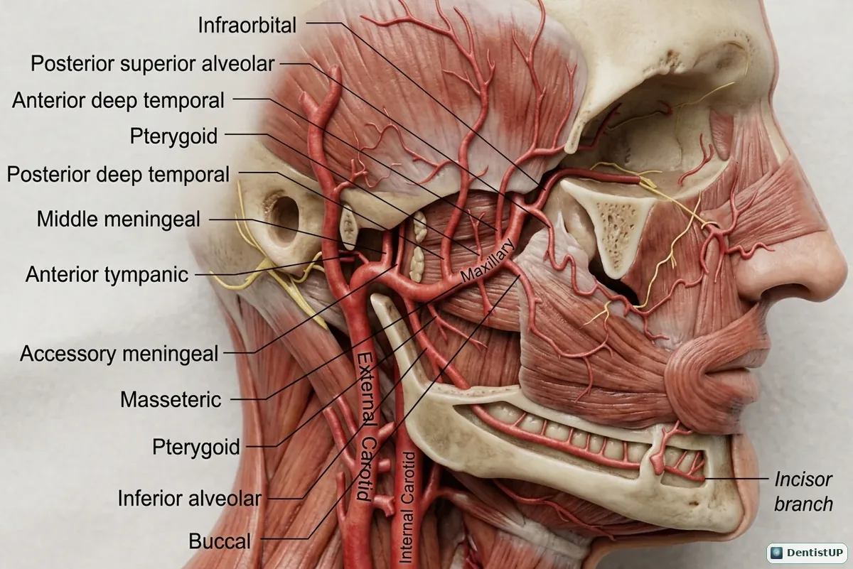
The maxillary artery runs through the infratemporal fossa into the pterygopalatine fossa. This illustration labels its branches, including the anterior tympanic, middle meningeal, accessory meningeal, inferior alveolar, deep temporal, masseteric, pterygoid, buccal, posterior superior alveolar and infraorbital arteries.
Exams divide the artery into three parts around the lateral pterygoid muscle and ask which branches arise from each. Clinically, the posterior superior alveolar vessels explain why a PSA block carries the highest risk of haematoma, and the inferior alveolar artery runs with its nerve through the mandibular canal. The middle meningeal artery, passing through the foramen spinosum, is the vessel injured in an extradural haematoma after a blow to the pterion.
Related practice questions
Educational illustration for exam preparation, not patient-specific clinical guidance.