
Paranasal Sinuses
Maxillary Sinus and Lateral Nasal Wall
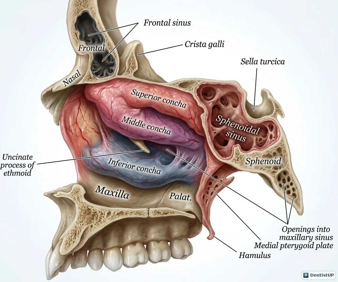
A sagittal view of the lateral nasal wall showing the superior, middle and inferior conchae, the frontal and sphenoidal sinuses, the uncinate process of the ethmoid, the medial pterygoid plate and hamulus, and the openings into the maxillary sinus beneath the middle concha.
The maxillary sinus drains into the middle meatus through an ostium high on its medial wall, so drainage depends on cilia rather than gravity. For dentists, the sinus floor lies close to the roots of the upper molars and premolars, which explains oroantral communication after extraction, root displacement into the sinus, odontogenic sinusitis, and the need for sinus augmentation before implant placement in the posterior maxilla.
Related practice questions
Educational illustration for exam preparation, not patient-specific clinical guidance.