
Paranasal Sinuses
Lateral Nasal Wall and Meatuses
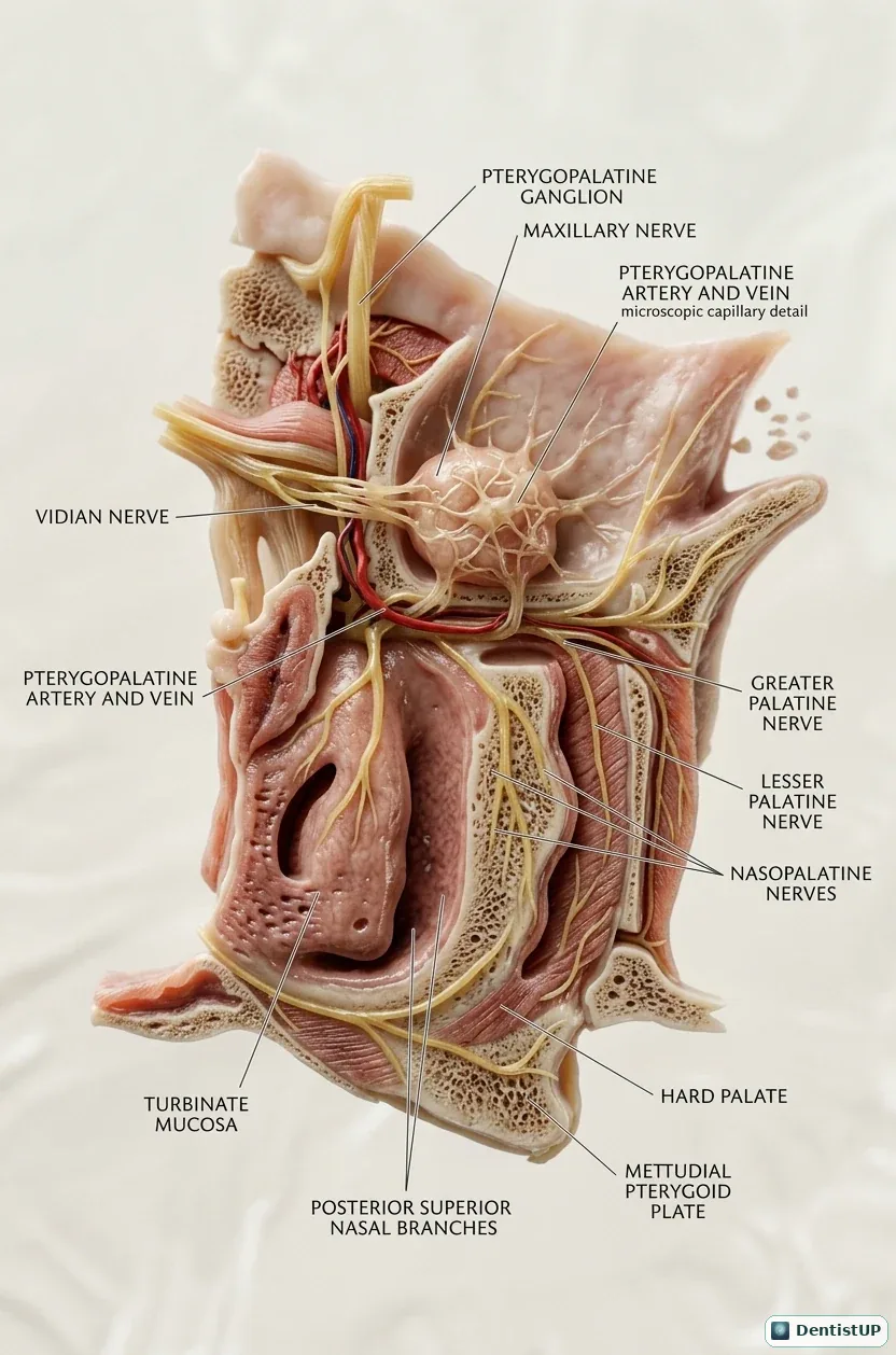
The lateral nasal wall with its conchae and meatuses, and the nerves and vessels reaching it from the pterygopalatine fossa: the pterygopalatine ganglion, the nerve of the pterygoid canal, the nasopalatine nerve, and the greater and lesser palatine nerves descending to the palate.
This anatomy explains palatal anaesthesia. The greater palatine nerve emerges at the greater palatine foramen, usually opposite the second or third molar, and supplies the posterior hard palate. The nasopalatine nerve runs across the septum to the incisive foramen and supplies the anterior palate. The pterygopalatine ganglion also carries secretomotor fibres to the lacrimal and nasal glands, which ties this diagram to the parasympathetic pathways of the head.
Related practice questions
Educational illustration for exam preparation, not patient-specific clinical guidance.