
Paranasal Sinuses
Paranasal Sinuses Overview
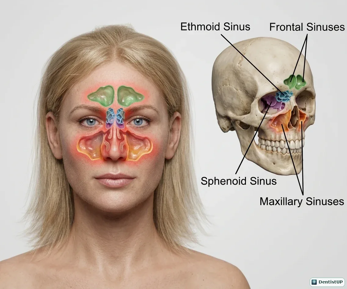
An overview of the four paired paranasal sinuses projected onto the face and skull: frontal, ethmoid, sphenoid and maxillary.
The maxillary sinus is the largest and the one dentists deal with most. It is present at birth but small, and it expands as the permanent teeth erupt, which is why its floor dips between the roots of the upper molars in adults. Sinus pain can mimic toothache in the upper posterior teeth, and a sinus infection can make several adjacent teeth tender to percussion at once, a pattern that helps separate sinusitis from a single-tooth endodontic problem. On panoramic radiographs the sinus floor and septa are common normal landmarks that candidates must not mistake for pathology.
Related practice questions
Educational illustration for exam preparation, not patient-specific clinical guidance.