
Skeletal Anatomy
Maxilla Bony Landmarks
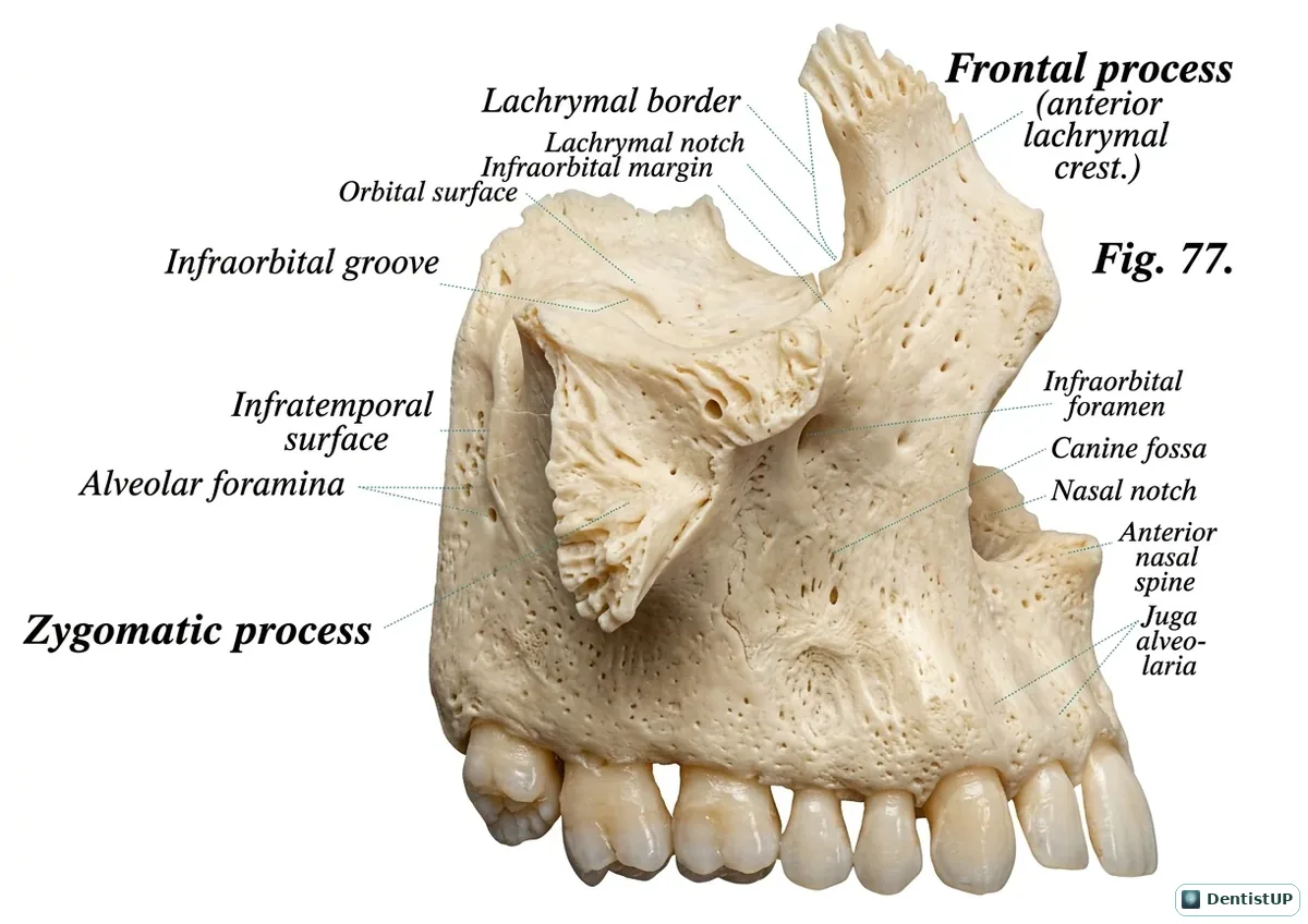
This view of the maxilla labels its frontal process, orbital surface and infraorbital margin, the infraorbital groove and foramen, the canine fossa, the zygomatic process, the infratemporal surface with its alveolar foramina, the nasal notch and anterior nasal spine, and the juga alveolaria over the tooth roots.
For dental board exams, the infraorbital foramen is the target of the infraorbital block and the exit of the infraorbital nerve, while the alveolar foramina on the infratemporal surface transmit the posterior superior alveolar nerves and vessels, which explains the positioning for a PSA block and the risk of haematoma. The canine fossa is a surgical access point to the maxillary sinus, and the thin bone over the roots explains why maxillary infections usually drain buccally.
Related practice questions
Educational illustration for exam preparation, not patient-specific clinical guidance.