
Cranial Foramina
External Skull Base Foramina
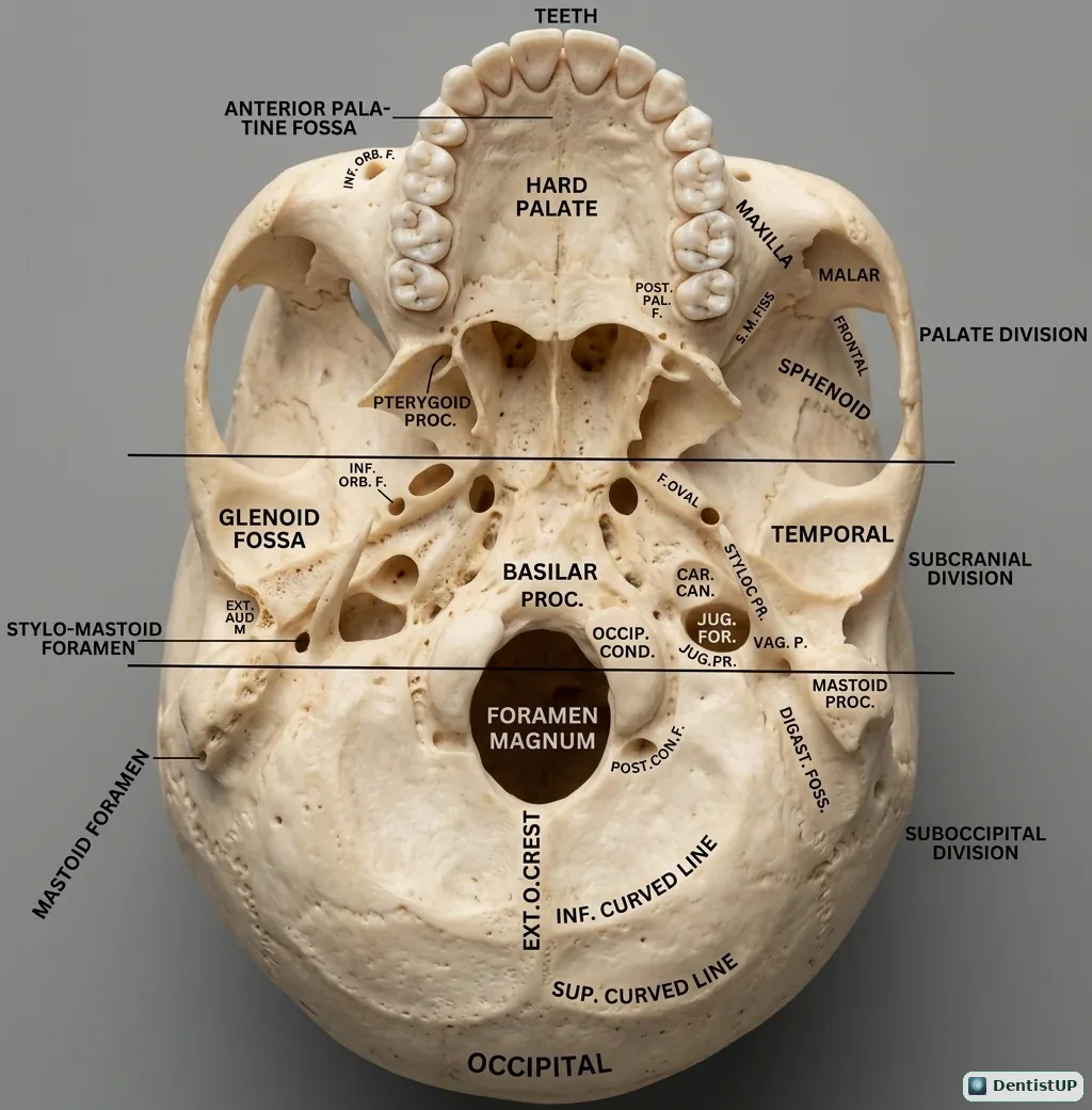
This inferior view of the skull base shows the hard palate and its foramina, the glenoid fossa, the stylomastoid foramen, the foramen magnum and occipital condyles, and the regions of the temporal and occipital bones.
Cranial foramina are a classic recall topic on the INBDE, AFK and ORE, and they are easiest to learn by position. The stylomastoid foramen transmits the facial nerve, which is why parotid surgery risks facial palsy. The foramen ovale, behind the lateral pterygoid plate, carries the mandibular division of the trigeminal nerve; the foramen spinosum carries the middle meningeal artery. Anteriorly, the incisive and greater palatine foramina are the landmarks for nasopalatine and greater palatine injections.
Related practice questions
Educational illustration for exam preparation, not patient-specific clinical guidance.