
Muscles
Muscles of Facial Expression
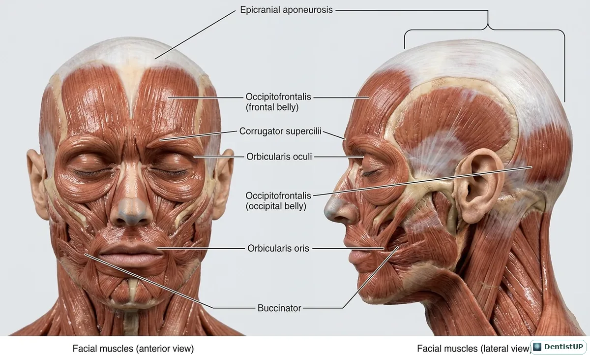
Anterior and lateral views of the muscles of facial expression, including the epicranial aponeurosis with the frontal and occipital bellies of occipitofrontalis, corrugator supercilii, orbicularis oculi, orbicularis oris and buccinator.
All of these muscles develop from the second pharyngeal arch and are supplied by the facial nerve, a link exam writers use often. Buccinator matters clinically: it keeps food on the occlusal table, is pierced by the parotid duct opposite the upper second molar, and its attachment determines whether a molar infection spreads into the buccal space or the vestibule. Loss of function in orbicularis oculi and oris after a misplaced inferior alveolar injection into the parotid is a recurring complication question.
Related practice questions
Educational illustration for exam preparation, not patient-specific clinical guidance.