
Muscles
Suprahyoid and Infrahyoid Muscles
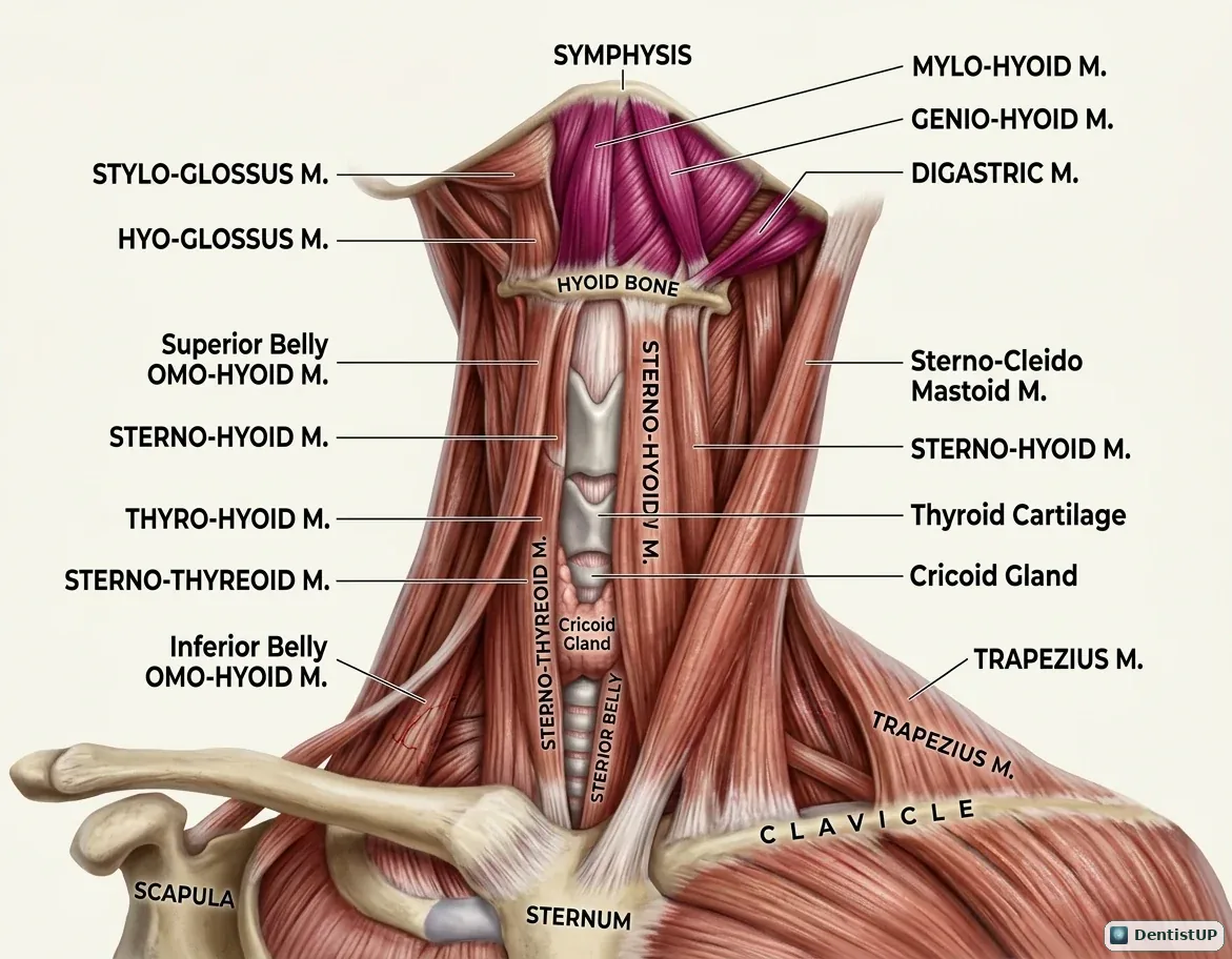
This illustration shows the muscles attached to the hyoid bone. Above it are mylohyoid, geniohyoid, the anterior and posterior bellies of digastric and stylohyoid; below it are sternohyoid, omohyoid, thyrohyoid and sternothyroid, with the thyroid cartilage and sternocleidomastoid for orientation.
The suprahyoid group raises the hyoid during swallowing and helps depress the mandible. Their innervation is a favourite exam item because it follows embryology: mylohyoid and the anterior digastric belly are supplied by the nerve to mylohyoid from V3, the posterior digastric belly and stylohyoid by the facial nerve, and geniohyoid by C1 fibres carried with the hypoglossal nerve. Mylohyoid also forms the floor of the mouth and separates the sublingual and submandibular spaces.
Related practice questions
Educational illustration for exam preparation, not patient-specific clinical guidance.