
Pharynx and Larynx
Larynx: Superior View
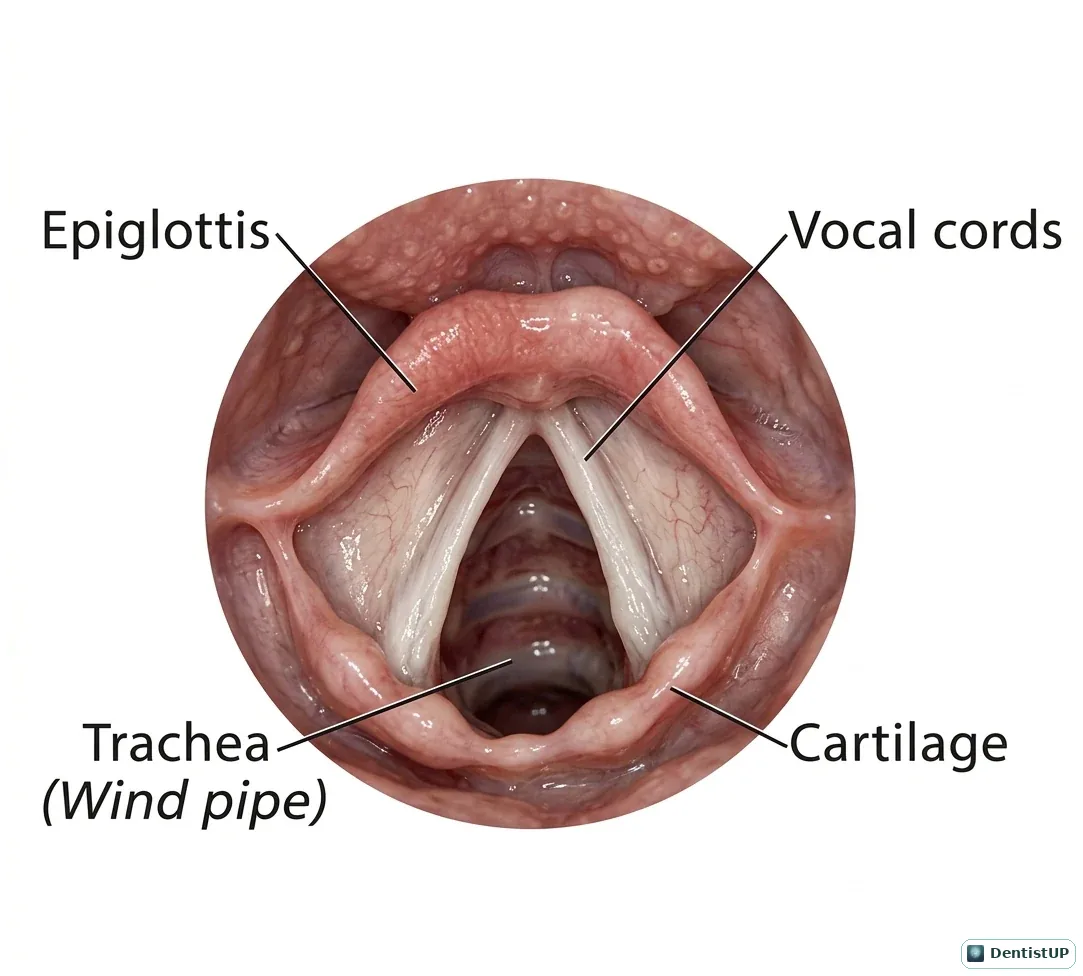
A view of the larynx from above, as seen during laryngoscopy, showing the epiglottis, the vocal cords forming the glottis, the surrounding cartilages and the trachea beyond.
Dentists do not intubate, but board exams expect you to understand the airway during sedation and medical emergencies. The glottis is the narrowest part of the adult airway, and laryngospasm, reflex closure of the vocal cords, is a recognised complication of sedation when blood or fluid reaches the larynx. Knowing the structures also helps with questions on aspiration of dental instruments and on the recurrent laryngeal nerve, which supplies all intrinsic laryngeal muscles except cricothyroid.
Related practice questions
Educational illustration for exam preparation, not patient-specific clinical guidance.