
Pharynx and Larynx
Pharynx Sagittal Anatomy
License this illustration for your lectures →
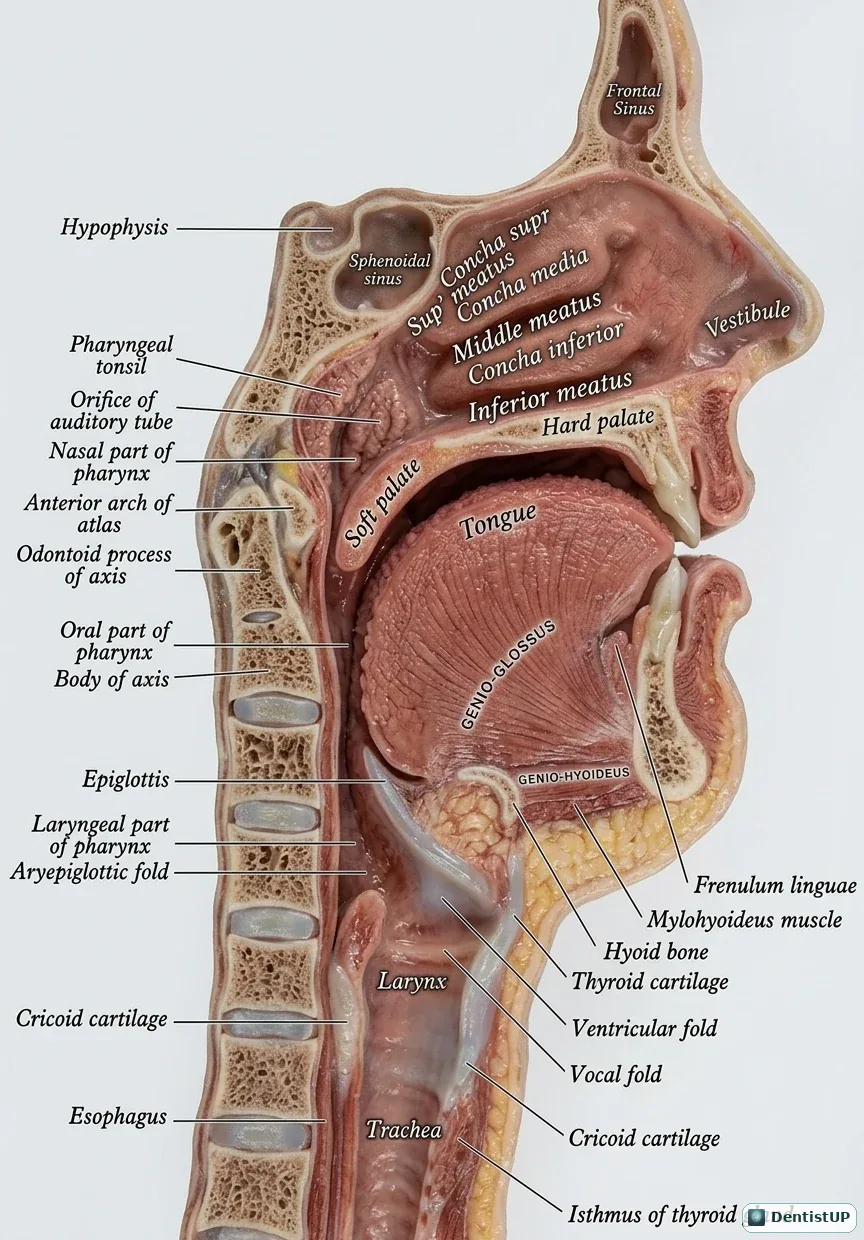
A midsagittal section showing the three parts of the pharynx: the nasopharynx with the opening of the auditory tube, the oropharynx behind the tongue and soft palate, and the laryngopharynx leading to the oesophagus. The larynx is shown with the epiglottis, vestibular and vocal folds, and the hyoid, thyroid and cricoid cartilages.
These relations matter for swallowing physiology, sedation and medical emergencies. During swallowing the soft palate closes off the nasopharynx and the larynx rises under the epiglottis. In an airway emergency, the cricothyroid membrane between the thyroid and cricoid cartilages is the site for emergency access. Aspirated objects in adults more often enter the right main bronchus, which is wider and more vertical.
Test yourself: 5 Head & Neck Anatomy questions
Free board-style questions with explanations. See all 10 →
Question 1 of 5
Paranasal Sinuses
The sphenoid sinus is located within which bone?
The answer is D.
The sphenoid sinus is located within the body of the sphenoid bone, positioned deep within the skull near midline structures such as the pituitary gland, internal carotid artery, and optic nerve, making its disease potentially serious.
Question 2 of 5
Muscles
The orbicularis oris muscle is primarily responsible for which action?
The answer is C.
The orbicularis oris is a circular sphincter muscle surrounding the mouth that closes and purses the lips, playing an important role in speech, feeding, and maintaining oral competence, and it receives its motor supply from the buccal and marginal mandibular branches of the facial nerve.
Question 3 of 5
Vasculature
The occipital artery, a branch of the external carotid artery, primarily supplies which region?
The answer is A.
The occipital artery branches from the external carotid artery and travels posteriorly to supply the scalp over the back of the skull, running a course that is relevant to surgical approaches involving the posterior neck and occipital region.
Question 4 of 5
TMJ
The stylomandibular ligament connects the styloid process to which part of the mandible?
The answer is D.
The stylomandibular ligament is a band of deep cervical fascia that extends from the styloid process of the temporal bone to the angle and posterior border of the mandibular ramus, helping to limit excessive protrusion of the mandible.
Question 5 of 5
Fascial Spaces
The submental space is located in which anatomic position?
The answer is A.
The submental space lies below the chin in the midline, bounded by the mylohyoid muscle above, the anterior bellies of the digastric muscles laterally, and the overlying skin and superficial fascia, and it is a common site of swelling from anterior mandibular incisor infections.
Choose an answer to reveal the explanation.
Related practice questions
Educational illustration for exam preparation, not patient-specific clinical guidance.