
Salivary Glands
Parotid Gland Microanatomy
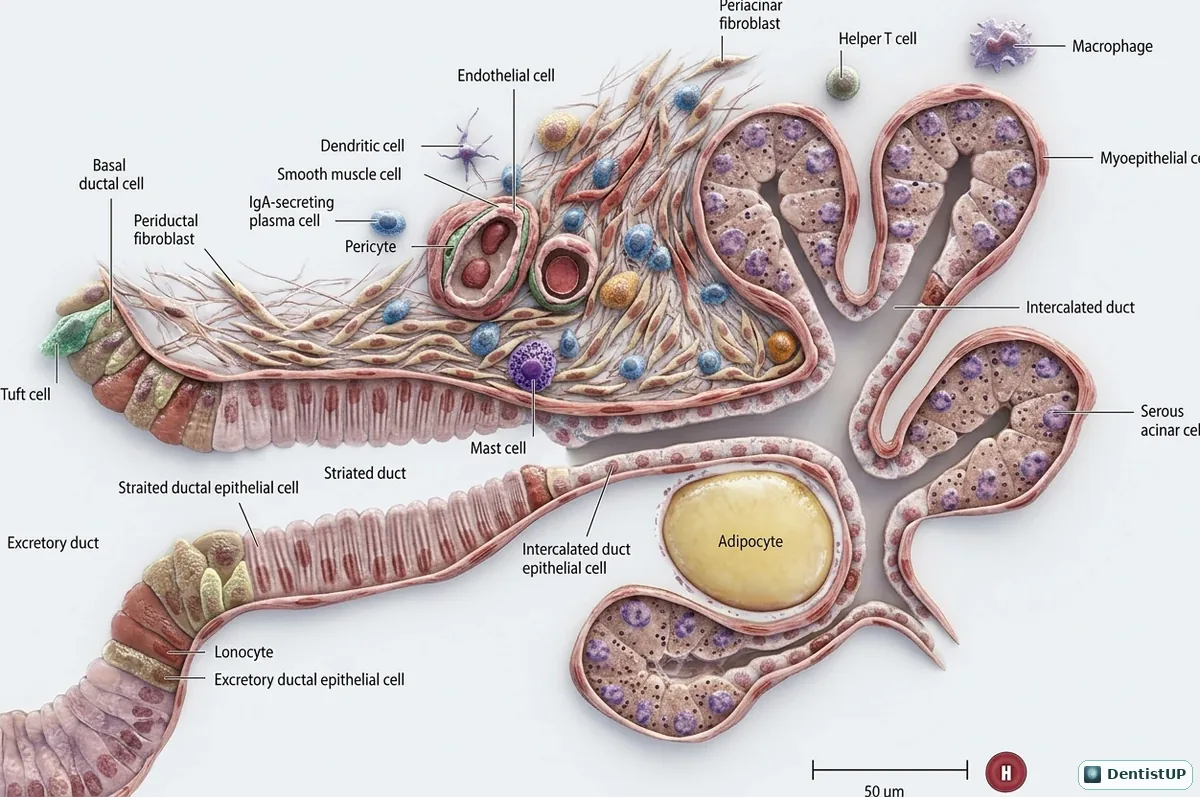
A microanatomical illustration of the parotid gland. Serous acini with myoepithelial cells drain into intercalated ducts, then striated ducts and finally excretory ducts. The surrounding stroma contains plasma cells, macrophages, mast cells, fibroblasts and adipocytes, with a scale bar for size.
The parotid is a purely serous gland in adults, which is the answer to a very common histology question, and it contains more fat with age. Striated ducts modify saliva by reabsorbing sodium, which is why saliva is hypotonic. Plasma cells in the stroma secrete IgA that is carried into saliva. Clinically, the parotid is the most common site of salivary tumours, and pleomorphic adenoma is the most common among them.
Related practice questions
Educational illustration for exam preparation, not patient-specific clinical guidance.