
Histology & Embryology Practice Questions
Oral histology and embryology form a core part of preclinical dental school and remain on board blueprints such as the INBDE and AFK. This set covers craniofacial embryology, odontogenesis, enamel, dentine, pulp, cementum, the periodontal ligament, oral mucosa, salivary gland histology and TMJ histology. Questions often ask which cells form a tissue, what happens at a given stage of tooth development, or how a developmental disturbance explains a clinical anomaly. Visual memory helps a great deal here, so working from clear diagrams of the bell stage, dentinal tubules and PDL fibre groups is worthwhile.
10 questions · answers and explanations revealed as you go · no sign-up
Questions
Question 1 of 10
TMJ Histology
A developing oral tissue shows dense avascular fibrous connective tissue between joint spaces. Which structure or stage is identified?
The answer is C.
Central articular disc is identified because it shows dense avascular fibrous connective tissue between joint spaces; its principal role is distribution of load and improvement of surface congruity. Its microscopic appearance and developmental role distinguish it from adjacent oral tissues.
Question 2 of 10
Salivary Gland Histology
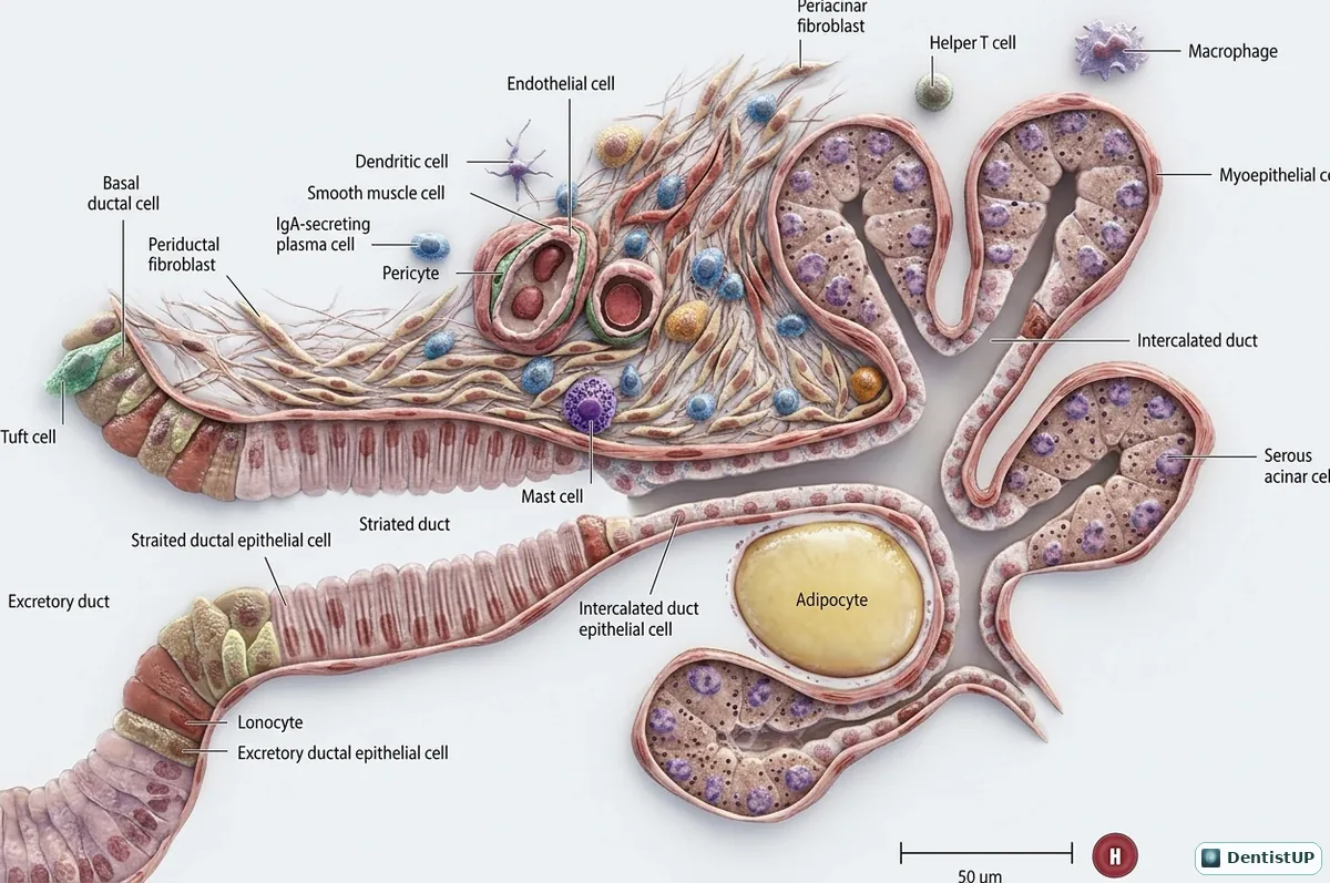
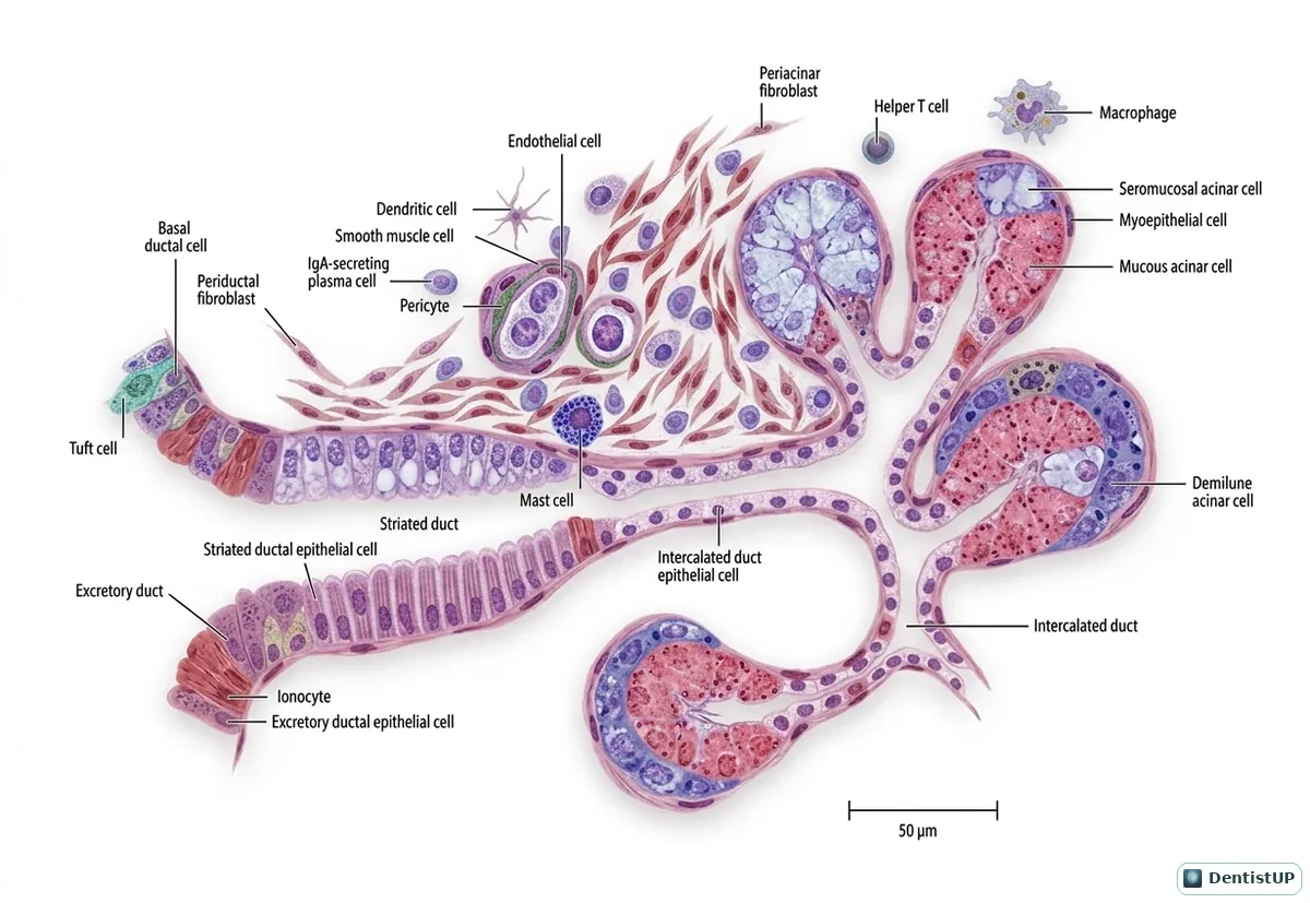
A developing oral tissue shows a stellate contractile cell between acinus and basal lamina. Which structure or stage is identified?
The answer is B.
Acinar myoepithelial cell is identified because it shows a stellate contractile cell between acinus and basal lamina; its principal role is expulsion of secretion from glandular endpieces. Its microscopic appearance and developmental role distinguish it from adjacent oral tissues.
Question 3 of 10
Cementum
A developing oral tissue shows rapidly formed apical cementum containing cementocytes. Which structure or stage is identified?
The answer is B.
Cellular intrinsic fiber cementum is identified because it shows rapidly formed apical cementum containing cementocytes; its principal role is adaptation and repair after root development. Its microscopic appearance and developmental role distinguish it from adjacent oral tissues.
Question 4 of 10
Pulp
A developing oral tissue shows fibrotic tissue in a narrowed pulp chamber. Which structure or stage is identified?
The answer is A.
Aged dental pulp is identified because it shows fibrotic tissue in a narrowed pulp chamber; its principal role is reduced reparative and sensory reserve over time. Its microscopic appearance and developmental role distinguish it from adjacent oral tissues.
Question 5 of 10
Odontogenesis
A developing oral tissue shows rounded epithelial buds entering ectomesenchyme. Which structure or stage is identified?
The answer is A.
Dental lamina epithelial ingrowth is identified because it shows rounded epithelial buds entering ectomesenchyme; its principal role is initiation of individual tooth germs. Its microscopic appearance and developmental role distinguish it from adjacent oral tissues.
Question 6 of 10
Enamel
A developing oral tissue shows a keyhole-like prism extending toward the surface. Which structure or stage is identified?
The answer is D.
Enamel rod is identified because it shows a keyhole-like prism extending toward the surface; its principal role is organization of crystals along a decussating path. Its microscopic appearance and developmental role distinguish it from adjacent oral tissues.
Question 7 of 10
Periodontal Ligament
A developing oral tissue shows small epithelial islands near the cementum surface. Which structure or stage is identified?
The answer is D.
Epithelial rests of Malassez is identified because it shows small epithelial islands near the cementum surface; its principal role is maintenance of ligament space and epithelial signaling. Its microscopic appearance and developmental role distinguish it from adjacent oral tissues.
Question 8 of 10
Oral Mucosa
A developing oral tissue shows flexible nonkeratinized stratified squamous epithelium. Which structure or stage is identified?
The answer is B.
Buccal lining mucosa is identified because it shows flexible nonkeratinized stratified squamous epithelium; its principal role is accommodation of cheek movement without surface cracking. Its microscopic appearance and developmental role distinguish it from adjacent oral tissues.
Question 9 of 10
Dentin
A developing oral tissue shows tertiary matrix made by replacement odontoblast-like cells. Which structure or stage is identified?
The answer is A.
Reparative dentin is identified because it shows tertiary matrix made by replacement odontoblast-like cells; its principal role is barrier formation after original odontoblast death. Its microscopic appearance and developmental role distinguish it from adjacent oral tissues.
Question 10 of 10
Craniofacial Embryology
A developing oral tissue shows an arch associated with glossopharyngeal nerve. Which structure or stage is identified?
The answer is C.
Third pharyngeal arch is identified because it shows an arch associated with glossopharyngeal nerve; its principal role is formation of stylopharyngeus and part of the hyoid. Its microscopic appearance and developmental role distinguish it from adjacent oral tissues.
Choose an answer to reveal the explanation.
Related anatomy diagrams
More branches
Full mock exams
DentistUP is an independent educational platform. Socket Science is not affiliated with, endorsed by, sponsored by, or approved by NDEB, INBDE, JCNDE, ADA, GDC, ORE, ADC, or any other examination authority. DentistUP does not contain official or recalled examination questions and does not guarantee examination outcomes. Educational content, not patient-specific clinical advice.