
Salivary Glands
Submandibular Gland Microanatomy
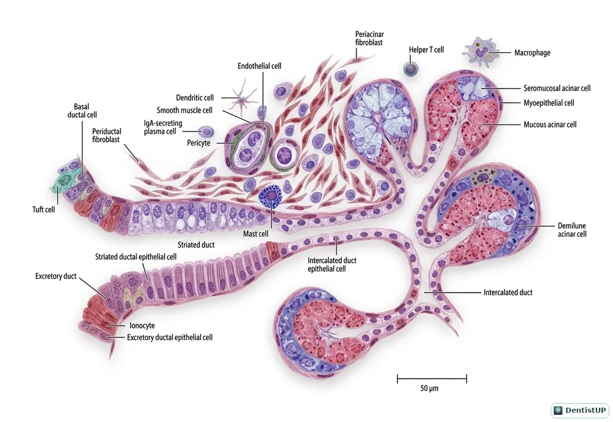
A microanatomical illustration of the submandibular gland showing serous and mucous acini, serous demilunes capping mucous acini, myoepithelial cells, and the duct system from intercalated to striated to excretory ducts.
The submandibular gland is mixed and predominantly serous, and it produces most unstimulated saliva. Serous demilunes are a classic histology image; in routinely processed tissue they are partly a fixation artefact. Clinically, the long, upward-running Wharton's duct and more alkaline, mucin-rich saliva explain why the submandibular gland is the most common site of salivary stones, a point that appears in both oral medicine and oral pathology questions. Sialadenitis and sialolithiasis of this gland typically cause swelling that worsens at mealtimes.
Related practice questions
Educational illustration for exam preparation, not patient-specific clinical guidance.