
TMJ
TMJ Labelled Anatomy
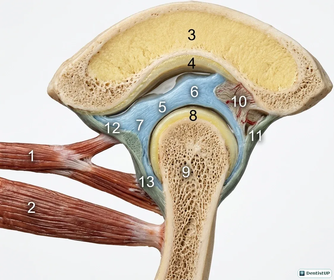
A numbered sagittal illustration of the temporomandibular joint for self-testing. The numbered structures include the mandibular condyle and neck, the articular disc with its thin intermediate zone, the upper and lower joint spaces, the bone of the mandibular fossa, the retrodiscal tissue behind the disc, and the heads of lateral pterygoid inserting in front.
Covering the labels and naming each numbered structure is a useful way to prepare for anatomy and TMD questions. Know that the intermediate zone is the thin, avascular part that bears load; that the retrodiscal tissue (bilaminar zone) is vascular and innervated, which explains pain when the disc displaces anteriorly; and that the articular surfaces are covered by fibrocartilage rather than hyaline cartilage, a frequent histology question.
Related practice questions
Educational illustration for exam preparation, not patient-specific clinical guidance.