
TMJ
TMJ Sagittal Anatomy
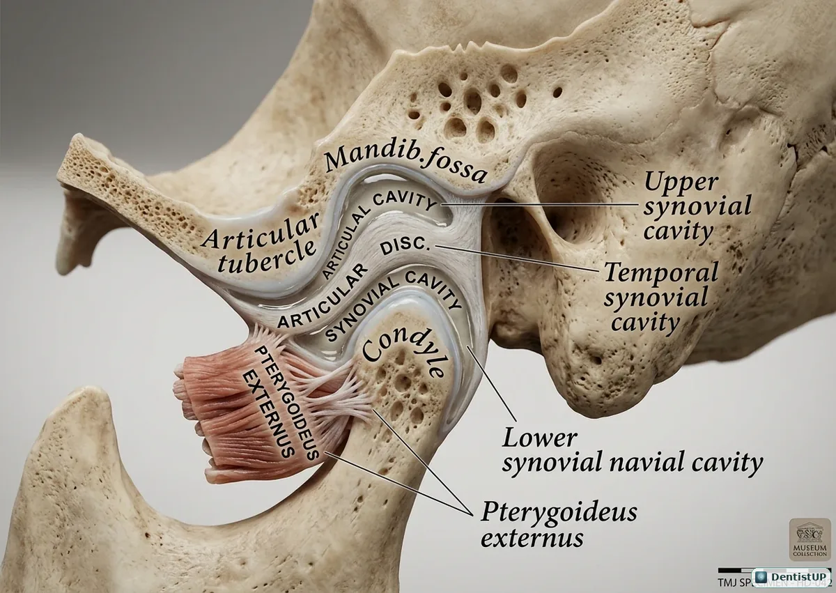
A sagittal section through the temporomandibular joint showing the mandibular (glenoid) fossa, the articular tubercle, the articular disc dividing the joint into upper and lower synovial cavities, the condyle, and the lateral pterygoid muscle attaching in front.
This view explains the two movements of the joint. Rotation happens in the lower compartment between the condyle and disc; translation happens in the upper compartment as the disc-condyle complex slides forward onto the articular eminence. Anterior disc displacement, with or without reduction, is described relative to this anatomy and is one of the most tested TMD topics. The lateral pterygoid's pull on the disc and condyle is central to both normal opening and dislocation.
Related practice questions
Educational illustration for exam preparation, not patient-specific clinical guidance.